
Human Eye and Defects is an important topic in Physics that explains how our eye works as a natural optical instrument. It also helps us understand common vision defects like myopia and hypermetropia, along with their corrections.
Previous year Questions
| Year | Question | Marks |
| 2021 | Describe the functioning of the human eye and explain any one of the refractive defects of vision and its corrective measure. | 10M |
| 2016 | What is hypermetropia ? How can this defect be corrected ? | 2M |
The Human Eye: Structure and Function
The human eye is one of the most important and sensitive sense organs, allowing us to perceive the colors, shapes, and details of the world around us. Color perception is only possible through vision.
The human eye functions like a camera, forming images on a light-sensitive surface known as the retina.
Structure of the Human Eye
The human eye is a nearly spherical organ with a diameter of about 2.3 cm. It is located in the eye socket (orbit) and protected by bones, eyelids, and tears. Light enters through various parts of the eye, which work together to create clear and focused images.

External Parts of the Eye
| Part | Function |
| Eyelids | Protect the eye from dust, bright light, and injury. |
| Eyelashes | Trap dust and small particles, preventing them from entering the eye. |
| Tear Glands (Lacrimal Glands) | Produce tears to keep the eye moist and wash away irritants. |
| Eyebrows | Prevent sweat and dust from falling into the eyes. |
Internal Parts of the Eye and Their Functions
| Part | Function |
| Cornea | A transparent, curved membrane at the front of the eye that refracts (bends) most of the light entering the eye. |
| Iris | A muscular diaphragm that controls the size of the pupil and, therefore, the amount of light entering the eye. The color of the eye is determined by the pigments in the iris. |
| Pupil | The black circular opening in the center of the iris that allows light to enter the inner parts of the eye. It dilates in dim light and contracts in bright light.This is why, when we move from bright light to dim light, our eyes cannot see properly for a while. After a short period, the size of the pupil increases, and we begin to see clearly. |
| Eye Lens | A flexible, transparent, biconvex structure that adjusts its shape to focus light on the retina for clear vision.The image formed is small, inverted, and real. |
| Ciliary Muscles | Muscles that control the shape of the lens by adjusting its curvature, allowing us to see objects at different distances. |
| Retina | A thin, light-sensitive membrane at the back of the eye, containing millions of photoreceptor cells (rods and cones)that convert light into electrical signals. |
| Optic Nerve | A bundle of nerve fibers that transmits visual information from the retina to the brain, where it is processed into meaningful images. |
| Aqueous Humor | A clear fluid between the cornea and the lens that provides nourishment and maintains the eye’s shape. |
| Vitreous Humor | A gel-like substance inside the eye that helps maintain its spherical shape and allows light to pass through. |
| Sclera | The white, tough outer layer of the eye that provides structure and protection. |
Working of the Human Eye
The eye works like a camera, capturing light and forming an image. The process includes:
Step 1: Light Enters the Eye
- Light enters through the cornea, which bends (refracts) it towards the pupil.
Step 2: Regulation of Light
- The iris adjusts the size of the pupil:
- Bright light → Pupil contracts to allow less light in.
- Dim light → Pupil expands to allow more light in.
Step 3: Focusing the Image
- The lens changes shape (accommodation) to focus light directly on the retina.
- The ciliary muscles help adjust the lens for clear vision at different distances:
- Looking at nearby objects → Lens becomes thicker.
- Looking at distant objects → Lens becomes thinner.
Step 4: Image Formation on the Retina
- The retina receives the light and forms an inverted, real image.
Step 5: Conversion of Light to Electrical Signals
- The retina contains photoreceptors that convert light into electrical signals.
- Rods – Help in night vision (detect black, white, and shades of gray).
- Cones – Detect colors (red, green, blue).
Step 6: Transmission to the Brain
- The optic nerve carries these signals to the brain, which processes them into an upright image.
Binocular Vision:
- Humans have two eyes that work together, allowing for depth perception and 3D vision.
- This helps us judge distances accurately (e.g., catching a ball).
Persistence of Vision
- The human eye retains an image for about 1/16th of a second after the object is removed.
- This is called persistence of vision and is used in movies and animations.
Power of Accommodation
The eye lens is made of a flexible, jelly-like material, allowing it to change shape and adjust focus. This ability is called accommodation.
When looking at distant objects:
- Ciliary muscles relax, making the lens thin (less curved).
- Focal length increases, allowing clear vision of far objects.
When looking at nearby objects:
- Ciliary muscles contract, making the lens thicker (more curved).
- Focal length decreases, allowing clear vision of close objects.
Limitations of Accommodation
- Near Point: The closest distance at which an object can be seen clearly (~25 cm for a normal eye).
- Objects held too close to the eyes appear blurred, causing strain.
- Far Point: The farthest distance at which the eye can see clearly (infinity for a normal eye).
- With age, the lens becomes less flexible, leading to presbyopia (difficulty in near vision).
- As people age, the eye lens may become cloudy or milky, leading to partial or complete vision loss. This condition is called cataract.
Defects of the Human Eye
Eye defects occur when the eye fails to focus light properly on the retina, leading to blurred or distorted vision. These defects can be caused by:
- Irregular shape of the eyeball
- Improper functioning of the lens
- Weakness of the ciliary muscles
- Aging and genetic factors
Common Eye Defects
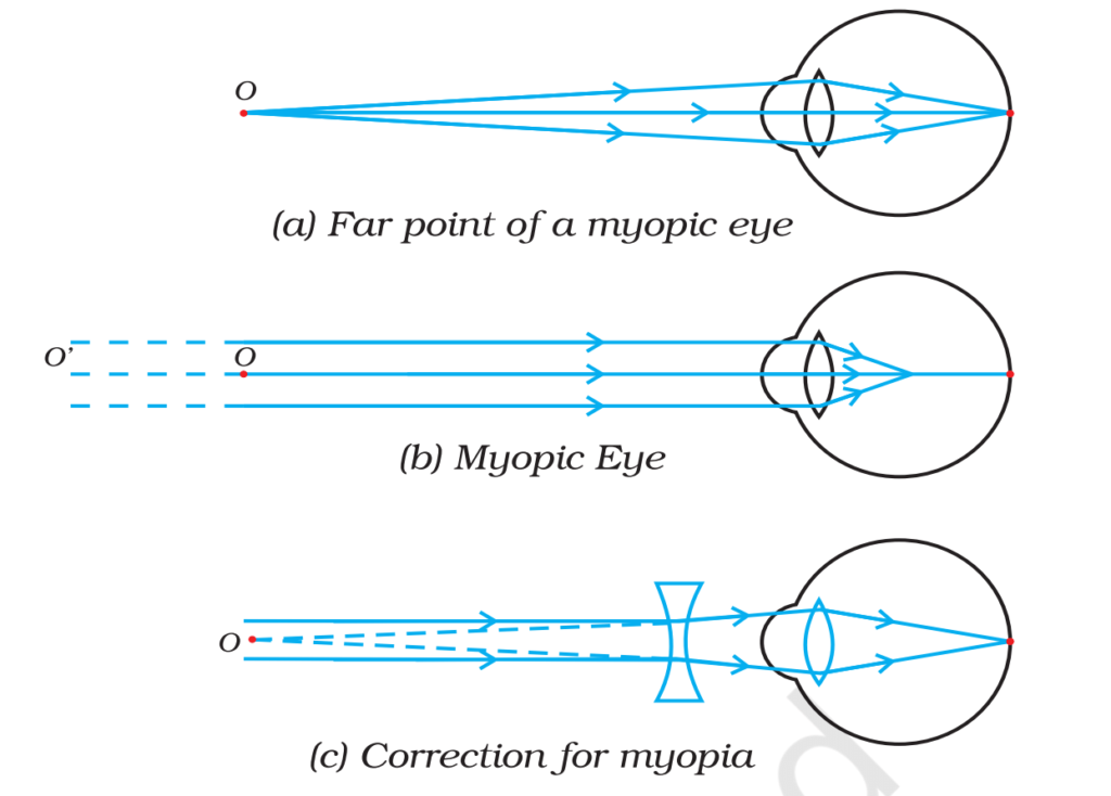
1. Myopia (Short-Sightedness or Near-Sightedness)
- Myopia is a refractive defect in which a person can see nearby objects clearly, but distant objects appear blurry.
Symptoms:
- Clear vision for nearby objects
- Blurred vision for distant objects
Cause:
- Excessive curvature of the eye lens (lens is too powerful).
- Elongation of the eyeball (light focuses before the retina instead of on it).
- A person suffering from this defect forms the image of distant objects before the retina, while the image of objects placed at a certain distance forms on the retina. In a way, the person’s far point is not at infinity, but rather it moves closer. This condition is known as myopia (nearsightedness).
Correction:
- Concave (Diverging) Lenses are used to spread out light before it enters the eye, so it focuses correctly on the retina.
- Alternative Treatments:
- Contact lenses (negative power).
- Laser surgery (LASIK) to reshape the cornea.
Example:
- A person with myopia struggles to read a board in class but can read a book easily.
2. Hypermetropia (Long-Sightedness or Far-Sightedness)
- Hypermetropia is a refractive defect in which a person can see distant objects clearly, but nearby objects appear blurry.
Symptoms:
- Clear vision for distant objects
- Blurred vision for nearby objects
Cause:
- The eyeball is too short, or the lens is too flat.
- Light focuses behind the retina instead of on it.
Correction:
- Using convex (converging) lenses, which bend light rays inward before they enter the eye.
- The convex lens brings the image forward onto the retina, restoring clear vision.
- Alternative Treatments:
- Contact lenses (positive power).
- Laser surgery to reshape the cornea and adjust the focal length.
Example:
- A person with hypermetropia struggles to read a book but can see distant objects clearly.

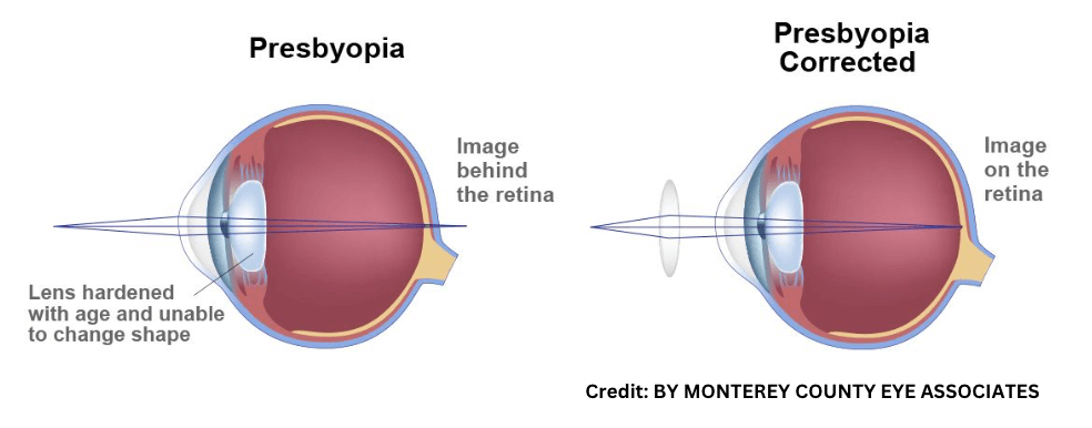
3. Presbyopia (Age-Related Long-Sightedness)
- Presbyopia is an age-related condition in which a person gradually loses the ability to see nearby objects clearly.
Symptoms:
- The near point recedes beyond 25 cm, making it hard to focus on close objects.
- People often need to hold books, phones, or newspapers farther away to read clearly.
Cause:
- The lens loses flexibility due to aging.
- Weakening of ciliary muscles (reduces ability to adjust lens curvature).
Correction:
- Convex lenses (like reading glasses) help focus on nearby objects.
- Bifocal lenses (a combination of concave & convex lenses) .
- Convex lenses help with near vision (lower part of the lens).
- Concave lenses help with distance vision (upper part of the lens).
- Alternative Treatments: Multifocal Contact lenses or surgical correction (e.g., LASIK) can also help.
Example:
Older people holding books at a distance to read clearly
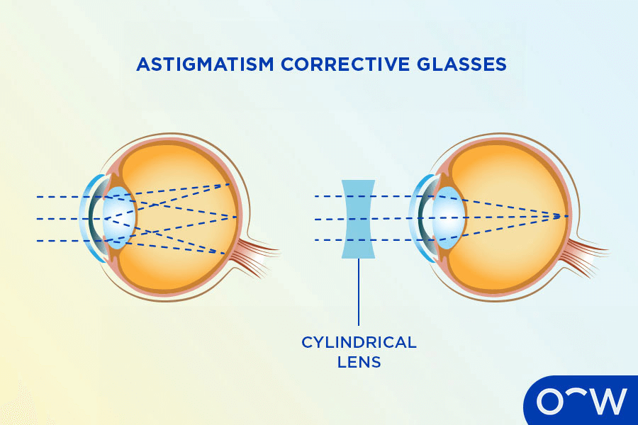
4. Astigmatism
Symptoms:
- Blurred or distorted vision at all distances.
- Difficulty seeing fine details.
Cause:
- The cornea is irregularly shaped, causing light to focus on multiple points instead of a single point.
Correction:
- Cylindrical Lenses correct the uneven curvature of the cornea.
Example:
- A person may see horizontal lines clearly but vertical lines appear blurry.
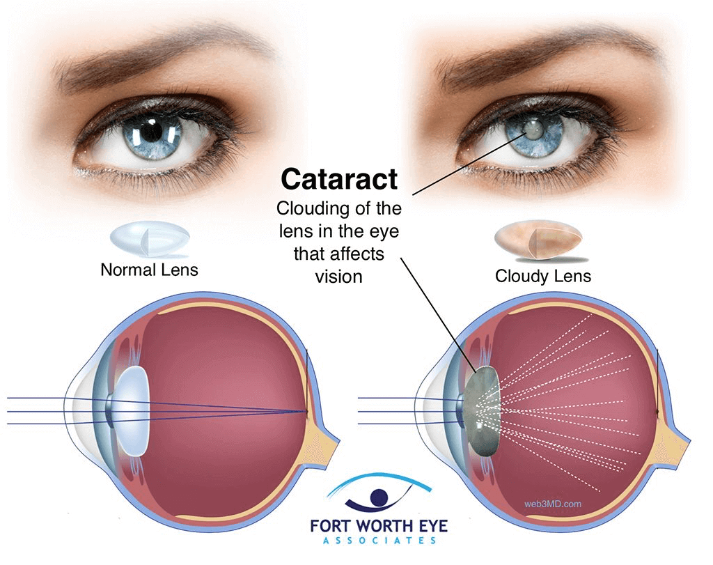
5. Cataract
As a person’s age increases, the transparency of the eye lens diminishes, and its flexibility decreases. Due to this, the lens starts reflecting light, causing objects to appear unclear. This condition is called a cataract.
Symptoms:
- Blurred or cloudy vision
- Sensitivity to bright light
- Faded colors
Cause:
- The lens becomes cloudy due to aging or injury, blocking light from entering the eye.
Correction:
- Surgical removal of the cloudy lens and replacement with an artificial lens.
Example:
- Elderly individuals who struggle with glare from headlights at night.
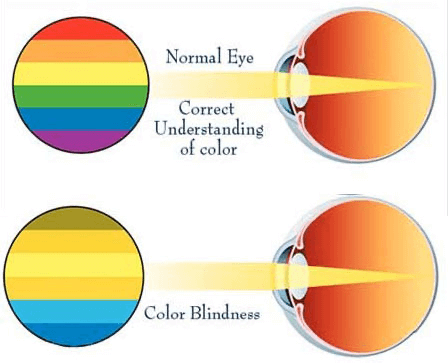
6. Color Blindness
Symptoms:
- Inability to distinguish certain colors (e.g., red and green).
Cause:
- Deficiency of cone cells in the retina (genetic condition).
Correction:
- No complete cure, but special tinted glasses can enhance color differentiation.
Example:
- A person with red-green color blindness may struggle to identify traffic lights correctly.
7. Night Blindness (Nyctalopia)
Cause:
- Deficiency of Vitamin A, which affects rod cells in the retina.
Symptoms:
- Difficulty seeing in low light or at night.
Correction:
- Vitamin A-rich diet (carrots, spinach, milk).
Example:
- A person struggles to see in dimly lit areas but has normal vision in daylight.
8. Photophobia (फोटोफोबिया)
- Photophobia refers to fear or discomfort in bright light. It is a condition where a person feels sensitive or in pain when exposed to intense light.
9. Xerophthalmia
- Xerophthalmia is a condition in which there is dryness in the eyes. It occurs due to a deficiency of Vitamin A in the body, which can lead to difficulty in producing tears and dry eyes.
Comparison of Common Eye Defects
| Defect | Cause | Effect on Vision | Correction |
| Myopia | Eyeball too long, lens too curved | Distant objects appear blurry | Concave lens |
| Hypermetropia | Eyeball too short, lens too flat | Nearby objects appear blurry | Convex lens |
| Presbyopia | Lens loses flexibility with age | Difficulty in near vision | Bifocal/progressive lenses |
| Astigmatism | Irregular cornea shape | Distorted vision at all distances | Cylindrical lens |
| Cataract | Clouding of lens | Blurred vision, glare sensitivity | Surgery |
| Color Blindness | Genetic defect in cone cells | Inability to differentiate colors | Special tinted glasses |
| Night Blindness | Vitamin A deficiency | Poor night vision | Vitamin A-rich diet |
FAQ (Previous year questions)
Functioning of the Human Eye-The human eye functions like a camera, capturing light and forming images.
works:
- Light Entry:Light first enters the eye through the cornea, which bends (refracts) it toward the pupil.
- Regulation of Light:The iris adjusts the size of the pupil:
- In bright light, the pupil contracts.
- In dim light, the pupil expands.
- Focusing the Image:The eye lens, with help from the ciliary muscles, changes its shape (accommodation) to focus light on the retina.
- For nearby objects, the lens becomes thicker.
- For distant objects, the lens becomes thinner.
- Image Formation: The retina receives the focused light and forms a small, inverted, real image.
- Conversion to Electrical Signals:The photoreceptor cells in the retina convert light into electrical signals:
- Rods help in dim light (black, white, grey).
- Cones detect colors (red, green, blue).
- Transmission to the Brain:The optic nerve transmits these signals to the brain, which interprets them as an upright, clear image.
- Binocular Vision: Both eyes work together to provide 3D vision and depth perception.
Refractive Defect: Myopia (Short-Sightedness)
- It is a condition where a person can see nearby objects clearly, but distant objects appear blurry.
- Cause:
- The eyeball is elongated, or
- The eye lens is too curved,
causing light rays from distant objects to focus in front of the retina instead of on it.
- Correction:
- Concave (diverging) lenses are used to spread out the incoming light rays so that they focus directly on the retina.
- Alternative treatments include contact lenses (with negative power) and LASIK surgery to reshape the cornea.
- Example:A student with myopia can read a book easily but has trouble seeing the classroom board clearly.
Hypermetropia (Long-Sightedness or Far-Sightedness)
- Hypermetropia is a refractive defect in which a person can see distant objects clearly, but nearby objects appear blurry.
Correction:
- Using convex (converging) lenses, which bend light rays inward before they enter the eye.
- The convex lens brings the image forward onto the retina, restoring clear vision.
- Alternative Treatments:
- Contact lenses (positive power).
- Laser surgery to reshape the cornea and adjust the focal length.
Functioning of the Human Eye-The human eye functions like a camera, capturing light and forming images.
works:
Light Entry:Light first enters the eye through the cornea, which bends (refracts) it toward the pupil.
Regulation of Light:The iris adjusts the size of the pupil:
In bright light, the pupil contracts.
In dim light, the pupil expands.
Focusing the Image:The eye lens, with help from the ciliary muscles, changes its shape (accommodation) to focus light on the retina.
For nearby objects, the lens becomes thicker.
For distant objects, the lens becomes thinner.
Image Formation: The retina receives the focused light and forms a small, inverted, real image.
Conversion to Electrical Signals:The photoreceptor cells in the retina convert light into electrical signals:
Rods help in dim light (black, white, grey).
Cones detect colors (red, green, blue).
Transmission to the Brain:The optic nerve transmits these signals to the brain, which interprets them as an upright, clear image.
Binocular Vision: Both eyes work together to provide 3D vision and depth perception.
Refractive Defect: Myopia (Short-Sightedness)
It is a condition where a person can see nearby objects clearly, but distant objects appear blurry.
Cause:
The eyeball is elongated, or
The eye lens is too curved,
causing light rays from distant objects to focus in front of the retina instead of on it.
Correction:
Concave (diverging) lenses are used to spread out the incoming light rays so that they focus directly on the retina.
Alternative treatments include contact lenses (with negative power) and LASIK surgery to reshape the cornea.
Example:A student with myopia can read a book easily but has trouble seeing the classroom board clearly.
Hypermetropia (Long-Sightedness or Far-Sightedness)
Hypermetropia is a refractive defect in which a person can see distant objects clearly, but nearby objects appear blurry.
Correction:
Using convex (converging) lenses, which bend light rays inward before they enter the eye.
The convex lens brings the image forward onto the retina, restoring clear vision.
Alternative Treatments:
Contact lenses (positive power).
Laser surgery to reshape the cornea and adjust the focal length.
