
Basics of Biology is a foundational topic in Science and Technology that helps us understand living organisms, their structure, functions, growth, and interactions with the environment. It covers essential concepts related to plants, animals, and the human body, building a strong base for further biological studies and real-life applications.
Plants
Nutrition in Plants
The process of intake of essential nutrients in the form of food for maintaining health, physical growth and development of an organism is called nutrition. Plants are the source of nutrients for all living beings The nutrients essential for the normal growth of plants which are absorbed from the soil can be mainly classified into two groups.
- Macro nutrients – those nutrients which are required by plants in large amounts. In plant tissues, their quantity is from 0.2% to 4%. Like carbon, hydrogen, oxygen, nitrogen, potassium, calcium, magnesium, sulphur etc.
- Further (a) Primary macro nutrients: nitrogen, phosphorus and potassium
- (b) Secondary macto nutrients: calcium, magnesium and sulphur.
- Micro nutrients– required in very low quantity for the healthy growth of plants. In plant tissues, their quantity is even less than 0.02% but still their presence is important for the plants. Zinc, copper, manganese, iron, boron, molybdenum, chlorine, nickel are placed in the category of micro nutrients. The deficiency of any of these nutrients causes diseases in plants.
Classification of plants on the basis of nutrition
- Parasite – obtain their food from other trees or plants are called Parasite whereas the tree or the plant from which the parasite obtains food is called the host. Example – Cuscuta
- Insectivorous plants – Those plants which in order to survive, trap insects and digest them.Example– drosera, dionaca, utricularia, pitcher plant etc.
- Saprotrophs – The plants which take nutrients from the dead and decaying matter. Example – Monotropa, Fungus on dung
- Symbiotic plants – Some organisms live together and share food, water, nutrients and shelter. Eg. lichens, two types of plants, fungi and an algae live together.
- Autotrophs – Those plants which prepare their own food with the help of sunlight.
Photosynthesis
- The process by which green plants use sunlight, water, and carbon dioxide to prepare carbohydrates (food) and release oxygen.
- Site of Food Synthesis – Food is synthesized in the leaves of plants. Leaves have small pores on their surface called stomata. Each stomatal pore is surrounded by guard cells.Carbon dioxide from air enters through stomata.
- Requirements for Photosynthesis To prepare food, leaves require: Sunlight, Water, Carbon dioxide, Mineral salts (brought from soil)
- Role of Roots and Stem – Roots absorb water and minerals from the soil. Stem contains tube-like vessels (xylem) that transport water and minerals to the leaves.
- Role of Chlorophyll – Leaves contain a green pigment called chlorophyll. Chlorophyll helps leaves capture sunlight energy. This energy is used to synthesize food.
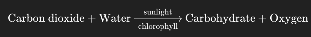
- End Products – Carbohydrates (used as food), Oxygen (released into the air), Carbohydrates are converted into starch and stored in the plant.
Following hormones are found in plants
Growth promoter
- Auxins: controls the growth of plants. Its main functions is to prevent the separation of the leaves.
- Gibberellins: It turns the dwarf plants into long plants. It helps in creating flowering. It helps in breaking the dormancy of plant.It motivates the seeds to sprout. It increases the activity of cambium in the wooden plants.
- Cytokinins– It helps in cell division and development, It helps in breaking the dormancy of seed. It is helpful in making RNA and protein.
Growth inhibitor
- Abscisic Acid or ABA : This hormone is against the growth. It keeps the seeds & bud in dormant condition. It plays the main role in separation of leaves. It delays the flowering of long day plants.
- Ethylene: It helps in the ripening of the fruits. It increases the number of female flowers. It motivates the separation of leaves, flowers and fruits. Gas used for artificial ripening of fruit is ethane or ethylene.
Other
- Florigens: It is formed in leaves but helps in blooming of the flowers. Therefore, it is also called flowering hormones.
- Traumatin: This is a type of dicarboxylic acid. It is formed in injured cells by which the injury of plants is healed.
Methods of asexual reproduction in plants
- Vegetative Reproduction : New plants grow from vegetative parts like root, stem, leaf, or bud. offspring are clones (identical to parent). Examples: Potato (eyes), Ginger, Turmeric (rhizomes), Bryophyllum (leaf buds), Rose, Money plant (stem cutting), Sweet potato, Dahlia (roots).
- Spore Formation : Reproduction by tiny spores with a hard coat → survive harsh conditions. Examples: Bread mould (Rhizopus),Ferns,Mosses.
- Fragmentation : Parent organism breaks into pieces, each piece grows into a new individual. Examples: Spirogyra (green algae), Other filamentous algae.
- Budding : A bud (small outgrowth) develops on the parent body, grows, and then detaches to form a new individual. Examples: Hydra (animal), Yeast (unicellular fungus).
System of the Human Body
Digestive System
The complete process of nutrition is divided into five stages : Ingestion, Digestion, Absorption, Assimilation, Defecation
- Ingestion : Taking the food into the mouth is called ingestion.
- Digestion : Conversion of nonabsorbable food into absorbable form. The digestion of the food starts from the mouth.
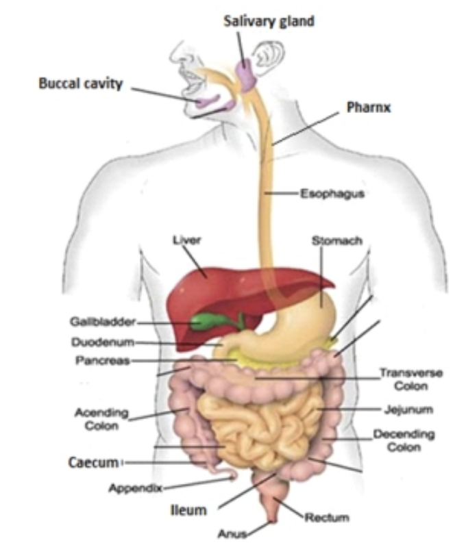
- Saliva is secreted by salivary gland in mouth in which enzyme amylase or ptyalin is present. They convert starch into simple sugar and make it digestible. The nature of saliva is acidic (pH 6.8). From the mouth food reach into stomach through food pipe or oesophagus. No digestion takes place in food pipe.
- The regular contraction and expansion of the wall of alimentary canal to push the food in forward direction is called peristaltic movement.
Digestion in Stomach
- After the food reaches the stomach, gastric glands secrete the gastric juice. pH of gastric juice is about 1.5 to 3.5.
- Hydrochloric acid secreted from the Oxyntic cells of the stomach kills all the bacteria coming with food and accelerates the reactions of enzymes. Hydrochloric acid makes the food acidic due to the action of salivary glands. The enzymes in the gastric juice of the stomach are – Pepsin and Rennin.
- Rennin breaks down the Caseinogen into Casein found in milk.
Digestion in Intestine
- Intestine is divided into two main part (a) Small intestine (b) Large intestine.
- Small intestine is sub divided into three part – Duodenum, Jejunum and Ileum.
Duodenum
- As soon as the food reaches the duodenum bile juice from liver combines with it. Bile juice is an alkaline and it turns the acidic medium of food into alkaline.
- Here, pancreatic juice from pancreas combines with food. It contains three types of enzymes :
- (a) Trypsin : It converts the protein and peptone into polypeptides and amino acid
- (b) Amylase : It converts the starch into soluble sugar.
- (c) Lipase : It converts the emulsified fats into glycerol and fatty acids.
- Cholecystokinin is a hormone secreted by endocrine cells of duodenum which induce the release of digestive enzyme from pancreas and bile from gall bladder. It is also known as appetite supressor.
Jejunum
- Here, the process of digestion completed and absorption of digested foods start.
- From the wall of small intestine, intestinal juices secretes. It contain following enzymes :
- (a) Erepsin : It converts the remaining protein and peptone into amino acids.
- (b) Maltase : It converts the maltose into glucose.
- (c) Sucrase : It converts the sucrose into glucose and fructose.
- (d) Lactase : It converts the lactose into glucose and galactose.
- (e) Lipase : It converts the emulsified fats into glycerol and fatty acids.
- Intestinal juice is alkaline in nature.
Ileum
- Absorption : The process by which digested food gets mixed into blood is called absorption. The absorption of digested foods takes place through villi found in the wall of the small intestinal or ileum.
- Assimilation : Use of absorbed food in the body is called assimilation.
Large intestine
- Defecation : Undigested food reaches into large intestine where bacteria turns it into faeces, which is excreted through anus.
Summary of Digestion
| Gland Juice | Enzyme | Edible Substance | After Reaction / Product |
| Saliva | Amylase | Starch | Maltose |
| Gastric Juice | Pepsin | Protein | Peptones |
| Rennin | Casein | Calcium paracasein | |
| Pancreatic Juice | Trypsin | Protein | Polypeptides |
| Amylase | Starch | Sugar | |
| Lipase | Fat | Fatty acid and glycerol | |
| Intestinal Juice | Erepsin | Protein | Amino acid |
| Maltase | Maltose | Glucose | |
| Lactase | Lactose | Glucose and fructose | |
| Sucrase | Sucrose | Glucose and galactose | |
| Lipase | Fat | Fatty acid and glycerol |
- Maximum digestion of food take place in Duodenum but complete digestion of food takes place in jejunum.
- Maximum absorption of food take place in jejunum but complete absorption of food take place in ileum.
The main organs participating in digestion :
Liver :
- This is the largest gland of the human body. Its weight is approximately 1.5–2 kilograms.
- It secretes Bile which emulsifies the fat in the intestine and facilitates the action of digestive enzymes.
- Liver converts excess amino acid into ammonia by deamination. Ammonia is further converted into urea by ornithine cycle. Urea is exerted out from the body through the kidney.
- In carbohydrates metabolism the liver converts the excess of glucose found in blood into glycogen and stores it into hepatic cell as reserve nutrients. If the necessity of glucose arises the liver converts glycogen into glucose. Thus, it regulates the quantity of glucose in the blood.
- In case of decrease of fat in food, the liver converts some of the parts of the carbohydrates into fat.
- The production of fibrinogen protein takes place by the liver which helps in clotting of blood.
- The production of Heparin protein takes place in the liver which prohibits the clotting of blood inside the body.
- Worn out or damaged RBC is destroyed by the liver only.
- Liver is an important clue in investigating a person’s death that has been due to poison in food.
Gall Bladder
- Gall bladder is a pear shaped sac, in which the bile coming from liver is stored.
- Bile comes into the duodenum from gall bladder through the bile duct.
- Secretion of bile into the duodenum takes place by reflex action. Bile is a yellowish-green coloured alkaline liquid. Its pH value is 7.7. The quantity of water is 85% and the quantity of bile pigment is 12%.
- The Main functions of bile are as under : It makes the medium of food alkaline so that pancreatic juice can worked. It kills the harmful bacteria coming with food. It emulsifies the fats. It accelerates the bowel movement of intestine by which digestive juices in the food mix well.
- In case of obstruction in bile duct, bile cannot be excreted in the intestine. As a result, it accumulates in the blood and spreads throughout the body. Causing jaundice.
Pancreas
- This is the second largest gland of the human body. It acts as simultaneously endocrine and exocrine type of gland.
- Pancreatic juice secretes out of it in which 9.8% water and the remaining parts contain salt and enzymes. It is alkaline liquid, whose pH value is 7.5–8.3.
- It contains the enzymes which can digest all the three types of food materials (like carbohydrates, fat and protein), therefore it is called complete digestive juice.
- Islets of Langerhans : This is a part of the Pancreas. From its β cell–insulin, from α cell–glucagons and from δ cell–somatostatin hormones are secreted.
- Glucagon : It re-converts the glycogen into glucose.
- Somatostatin : This is a polypeptide hormone which increases the duration of assimilation of food.
Respiratory System
- The most important organ of the human respiratory system is lungs where the exchange of gases takes place.
- The process of respiration is an exothermic oxidation reaction, because it releases energy in the form of heat and light, which fuels cellular activities.
- In humans, two lungs are situated in the thoracic cavity near the heart. The right lung has three lobes and the left lung has two lobes. Lungs are covered by a double layered membrane called the pleura.
- All those organs that come under the respiratory system are – Nasal passage, Pharynx, Larynx or Voice box, Trachea, Bronchi, Bronchioles, Lungs etc.
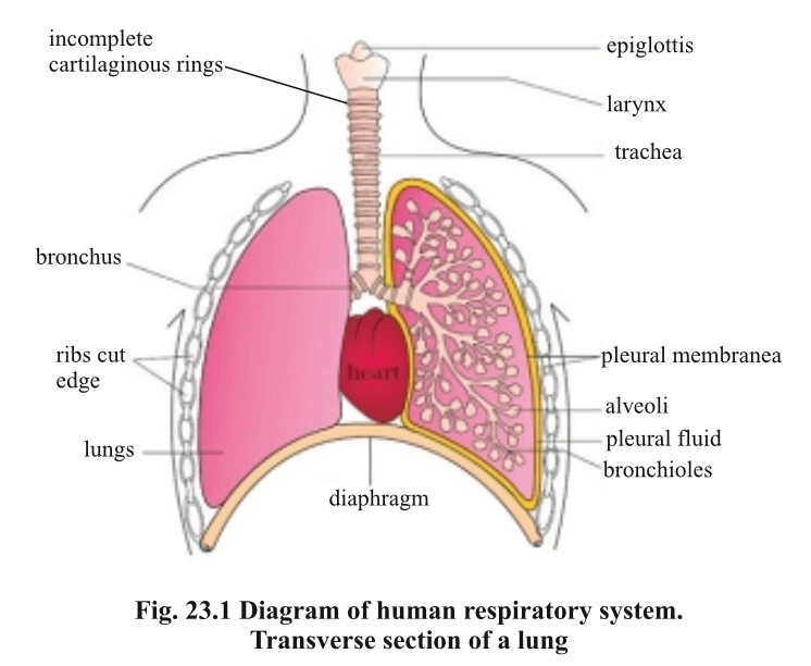
- Nasal Passage – It helps in breathing and smelling. The inner lining produces mucus which traps dust and germs. It also warms and moistens the air before it enters the lungs.
- Pharynx – It is a common passage for both air (respiration) and food (digestion). It lies behind the nasal cavity.
- Larynx – It is the voice box. The opening is called glottis and it is covered by epiglottis, which prevents food from entering the windpipe. Vocal cords present here help in producing sound.
- Trachea – It is a tube from the larynx to the chest. It is supported by C-shaped cartilage rings. Its inner lining has cilia and mucus to trap dust.
- Bronchi and Bronchioles – The trachea divides into two bronchi. Each bronchus enters a lung and further divides into tiny tubes called bronchioles. Bronchioles end in alveoli where gas exchange occurs.
- Lungs : There is a pair of lungs in the thoracic cavity. Its colour is pink, red and looks like sponge. Right lung is larger in comparison to left lung. Each lung is surrounded by a membrane which is called pleural membrane. There is a network of blood capillaries. Here oxygen enters into the blood and CO₂ release out from blood.
- The process of respiration can be divided into four parts—
- External respiration.
- Transportation of gases.
- Internal respiration.
- Cellular respiration.
1. External respiration :
- This is divided into two parts – (a) Breathing (b) Exchange of gases.
- (a) Breathing : In lungs air is taken and given out at a certain rate which is called breathing. An adult breath approximately 16 to 18 times in a minute.
Mechanism of Breathing :
- Inhalation is the entry of air into the body. It is an active phase which starts with the contraction of diaphragm and intercostal muscles. When the diaphragm contracts it becomes flat. At the time of contraction, diaphragm falls down in the abdomen, this increases the volume of thoracic cavity.
- Exhalation – The process of expelling air out of the lungs. In normal breathing it is a passive phase, whereas in forced breathing it becomes active.
- When external inter-costal muscles and muscles of diaphragm get relax then the ribs come down due to their own weight and the diaphragm rises up in thoracic cavity.
Constitution of air in Breathing
| Nitrogen | Oxygen | CO2 | |
| The air inhaled | 79% | 21% | 0.03% |
| The air exhaled | 79% | 17% | 4% |
Exchange of gases :
- The exchange of gases takes place inside the lungs. This gaseous exchange takes place on the basis of concentration gradient through normal diffusion.
- The exchange of oxygen and carbon dioxide gases takes place due to their difference in partial pressures. The direction of diffusion is both sides.
- Diaphragm is arched during normal expiration.
2. Transportation of gases :
- The process of reaching of gases (oxygen and carbon dioxide) from lungs to the cells of body and coming back again to the lungs is called the transportation of gases.
- Transportation of oxygen takes place by haemoglobin present in blood.
- Transportation of carbon dioxide from cells to lungs takes place by haemoglobin only to the extent of 10 to 20%.
- Carbon dioxide is transported through blood in three forms
- About 7% is transported dissolved in plasma.
- About 70% is transported in the form of bicarbonate ions.
- About 23% is transported by combining with hemoglobin as carbaminohaemoglobin.
3. Internal respiration :
- Inside the body, gaseous exchange takes place between blood and tissue fluid which is called internal respiration.
- Note : The gaseous exchange in lungs is called external respiration.
4. Cellular respiration :
- Cellular respiration is the process in which glucose is broken down inside the cell using oxygen to release energy needed for growth, repair, and daily activities.
- Types of cellular respiration : There are two types of Respiration
Anaerobic respiration :
- When the oxidization of food takes place in the absence of oxygen. During this only 2 ATP molecules are produced from one molecule of glucose.
- In animal tissues like skeletal muscle cells, anaerobic respiration produces lactic acid as the final product.
- In yeast and certain bacteria ethyl alcohol or ethanol is produced.
- C₆H₁₂O₆ → 2C₃H₆O₃(Lactic acid) + Energy (in animal)
- C₆H₁₂O₆ → 2C₂H₅OH (Ethyl alcohol) + 2CO₂ + Energy (in plant)
Aerobic respiration :
- It takes place in the presence of oxygen. The complete oxidation of glucose takes place. As a result CO₂ and H₂O is formed and energy is released in huge amount.
- C₆H₁₂O₆ + 6O₂ → 6CO₂ + 6H₂O + 2870 KJ energy. (38 ATP)
Respiratory substances :
- Respiratory substances are materials used by cells to release energy.
- Carbohydrates, fats, and proteins are the main respiratory substances.
- First glucose is oxidized, then fats, and finally proteins when others are exhausted.
Note :
- Respiration is a Catabolic process. It also reduces the weight of the body.
- Respiration is controlled by medulla oblongata.
- Cyanide poisoning causes death in seconds due to breaking down the electron transport chain system.
Endocrine System
- Exocrine glands : Glands which have ducts are called exocrine glands. They secrete enzymes or other substances that pass through these ducts. Example – Digestive gland, Sweat gland, Mucous gland, Salivary gland etc.
- Endocrine gland : These are ductless gland. Hormones are secreted by these gland. Hormones are sent to the different parts of the body through blood plasma. Example – Pituitary gland, Thyroid gland, Parathyroid gland etc.
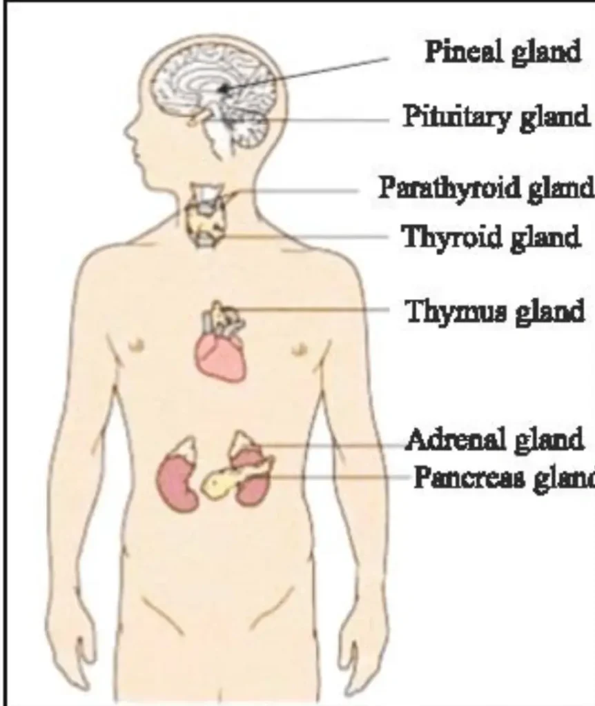
Pituitary gland :
- It is situated in a depression of the sphenoid bone of the fore head. This is also known as the master gland. The pituitary gland is controlled by hypothalamus. Hypothalamus is the part of the brain which controls emotional reaction in our body.
- The functions of the hormones secreted by the Pituitary gland, are as follows –
| Hormone | Function |
| Growth Hormone | Controls growth of body, especially bones |
| TSH (Thyroid Stimulating Hormone) | Stimulates thyroid gland to secrete hormone |
| ACTH (Adreno Corticotropic Hormone) | Controls secretion of adrenal cortex |
| FSH (Follicle Stimulating Hormone) | In males: stimulates spermatogenesis; In females: stimulates Graafian follicles to secrete estrogen |
| LH (Luteinizing Hormone) | In males: secretion of testosterone; In females: secretion of estrogen |
| Prolactin (Lactogenic Hormone) | Stimulates milk secretion in breasts |
| ADH (Antidiuretic Hormone) | Increases blood pressure, maintains water balance, reduces urine volume |
| Oxytocin | Helps uterine contraction during childbirth, releases milk from mammary glands (secreted by hypothalamus) |
Thyroid gland :
- This is situated below the larynx on both sides of the respiratory trachea in the throat of humans. The hormones secreted by it are Thyroxine and Triiodothyronine.
- Functions of Thyroxin :
- It increases the speed of cellular respiration. It is necessary for the normal growth of the body particularly for the development of bones, hair etc.
- The normal functions of reproductive organs depend on the activeness of the thyroid gland.
- It controls the water balance of the body in coordination with the hormones of the pituitary gland.
- Diseases Caused by the Deficiency of Thyroxin :
- Cretinism : This disease affects the children. The mental and physical retardness of the child.
- Myxedema : In this disease which normally attack during youth the metabolism does not take place properly which causes reduction in heart beat and blood pressure.
- Hypothyroidism : This disease is caused due to a chronic deficiency of thyroxin hormone. Due to this disease normal reproduction is not possible. Sometimes due to this disease a person becomes dumb and deaf.
- Goitre : This disease is caused by the deficiency of iodine in food. In this disease the shape of the thyroid gland enlarges abnormally. Iodization of salt is a public health measure to prevent goitre.
Parathyroid gland :
- This is situated in the right back of the thyroid gland of the throat. Two hormones are secreted by it :
- (a) Parathyroid hormone : This hormone is secreted when there is a deficiency of calcium in the blood.
- (b) Calcitonin : This hormone is released when there is excess of calcium in the blood is present.
- Hence, hormones secreted by the parathyroid gland controls the quantity of calcium in the blood.
Adrenal gland :
- There are two parts of this gland— (a) outer part is cortex and (b) inner part is medulla.
- Hormones secreted by cortex and their function—
- (i) Glucocorticoids : This controls the metabolism of carbohydrate, protein and fat.
- (ii) Mineralocorticoids : Its main function is reabsorption of ions by kidney ducts and to control the quantity of other ions in the body.
- (iii) Sex hormone : It controls the sexual behaviour and secondary sexual characters.
- Note : In case of deformation of cortex, the process of metabolism gets disturbed; this disease is called Addison’s disease.
Adrenaline and noradrenaline hormone
- They are secreted from the medulla of the adrenal gland during emergency conditions.
- They are secreted less in normal conditions more in stressful situations that’s why they are called stress hormone or emergency hormone.
- During their secretion heart rate, blood rate, respiration rate and sweating increase.
- Blood flow to the skeletal muscles increases and they make a person ready for fight or flight in emergency conditions so that’s why they are called 3F (Fear,Fight,Flight) hormone.
- When adrenaline is secreted, the body prioritizes blood flow to vital organs like the heart, brain, and muscles. This means that blood flow to less critical areas like the digestive system and skin is reduced.
- Thymus is an endocrine gland situated in the chest cavity and produces the hormone thymosin.
Gonads :
- Ovary : The following hormones are secreted by this
- (a) Estrogen : It completes the development of reproductive organs.
- (b) Progesterone : It stimulates the thickening of uterus lining during ovarian cycle.
- (c) Relaxin : During pregnancy it is found in uterus and placenta. This hormone smoothens the pubic symphysis and it widens the uterine cervix so that a child is delivered easily.
- Oxytocin is a hormone which helps in contraction of uterine wall during child birth and plays an important role in release of milk from the mammary glands secreted by hypothalamus.
- Testes : The hormone secreted by it is called testosterone. It motivates the sexual behaviour and growth of secondary sexual characters.
- Oxytocin is a hormone secreted by the posterior pituitary gland that causes contraction of the uterus during childbirth.
Excretory System
- In humans kidneys are main excretory organs. Besides kidneys, ureter, urinary bladder, Urethra also takes part in the excretion process.
- Excretion : Removal of nitrogenous substances formed during metabolism from the body of human is called excretion. Normally excretion means the release of nitrogenous excretory substances like urea, ammonia, uric acid etc.
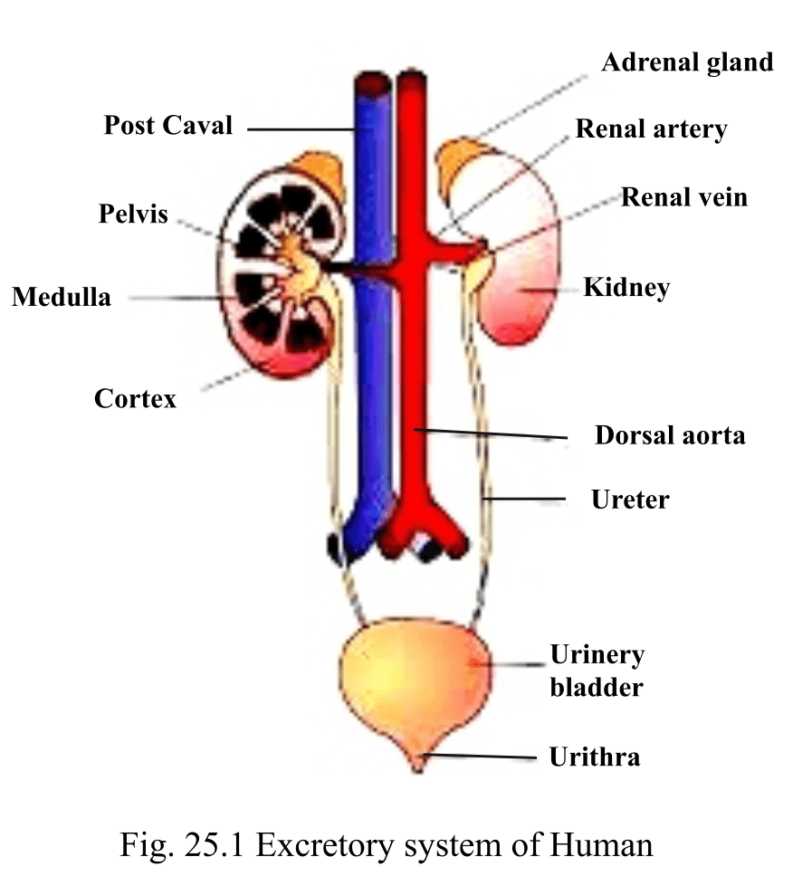
Kidneys :
- The main excretory organ in humans and other mammals is a pair of kidneys.
- There are two parts of it. The outer part is called cortex and the inner part is called medulla. Each kidney is made up of approximately 1,30,0000 kidney ducts which are called nephrons.
- Nephron is the structural and functional unit of the kidney.
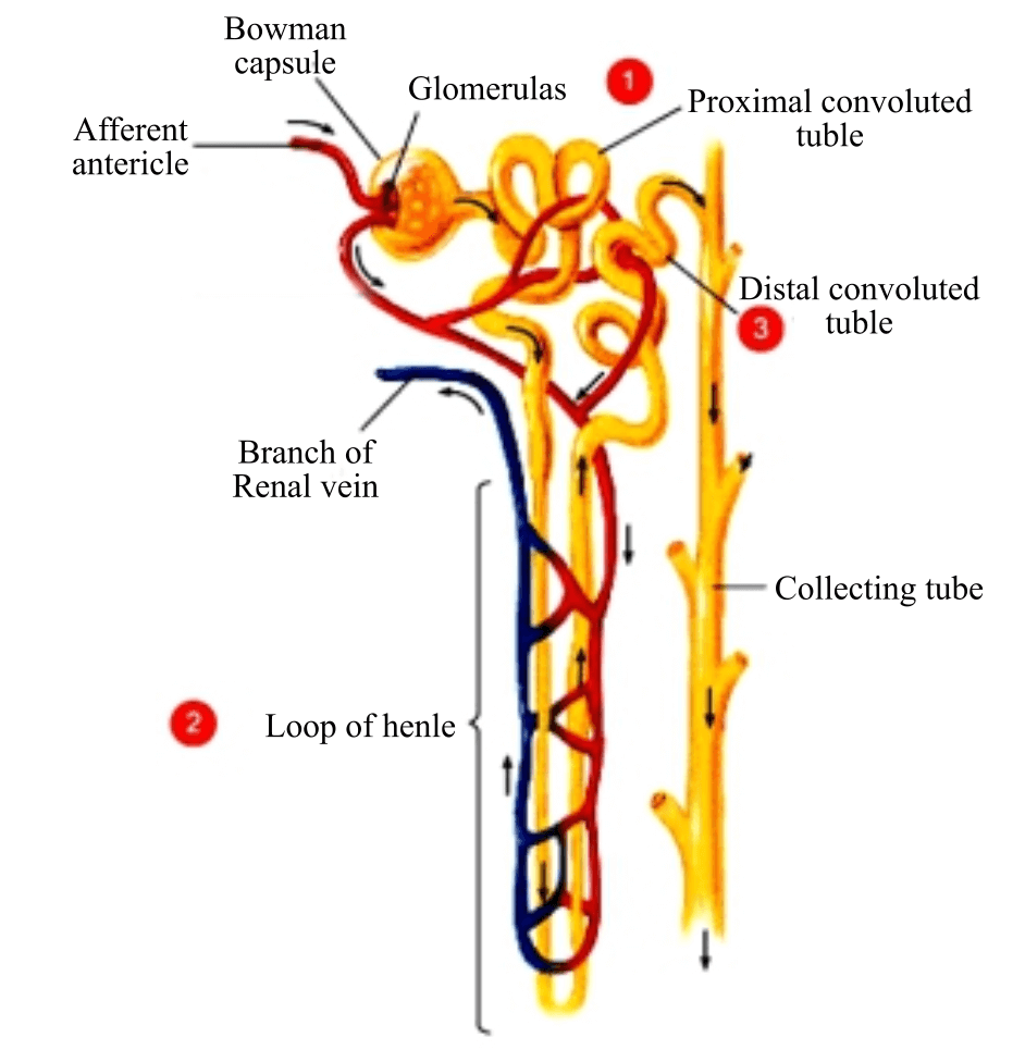
- There is a cup-like structure in every nephron called Bowman’s capsule. Glomerulus is made up of thin blood vessels found in the Bowman’s capsule.
- The process of filtration of liquids into the cavity of Bowman’s capsule, is called ultra filtration.
- The main function of the kidneys is purification of blood plasma i.e. to excrete the unwanted nitrogenous waste substances through urination.
- The supply of blood to kidneys takes place in large quantity in comparison to other organs.
- Urine is an aqueous solution in which 95% is water. Other constituents are urea, uric acid, chloride, sodium, potassium, creatinine, organic and inorganic compound.
- Urine Formation process
- Ultra filtration takes place in Bowman’s capsule – small size useful and waste product came into nephron from blood
- Reabsorption – small size useful matter again came back into blood. From this process takes place in the whole nephron but maximum in the proximal convoluted tube.
- Secretion – Large size useless matter enter Into urine from blood
- Conversion of ammonia into urea is called urea cycle which takes place in liver.Its first product is ornithine so also called the ornithine cycle.
- The colour of the urine is light yellow due to the presence of urochromes in it. Urochromes are formed by the dissociation of haemoglobin. Urine is acidic as its pH value is 6.
- The stones formed in the kidneys are made up of calcium oxalate.
- Other excretory organs in humans
- Skin – nitrogenous substances are excreted in the form of sweat.
- Liver : Liver cells play the main role in excretion by converting more and more amino acids and ammonia of blood into urea.
- Lungs : The lungs excretes two types of gaseous substances: carbon dioxide and water vapour. The Lungs excrete vapor components of some substances like garlic, onion.
Excretory disorder
- Uremia: When the blood amount of urea increases from 10-30 mg/100 ml then the state is known as uremia.
- Gout: This is a hereditary disease in which uric acid increases in blood which gets collected in joints and kidney tissues. This disease increases due to dehydration, fast and diuretic.
- Kidney stones: Commonly, crystals of uric acid, calcium oxalate, phosphate salts etc. deposit into renal pelvis in the form of stones. This creates pain in patients and obstruction in passing urine.
- Bright’s disease or nephritis: This disease is caused by the infection of bacteria – Streptococci in glomerulus. Due to this glomerulus develops inflammation and its membranes become too permeable so erythrocytes (RBC) and protein also sieve out in the filtrate.
- Diabetes Insipidus – Hypo secretion of antidiuretic hormone (ADH) develops checks on the absorption of water. This increases the volume of urine and the patient does urination again and again in increased volume.
- Jaundice: Presence of excessive bile pigments in urine is called jaundice. This is seen normally in hepatitis or blocked bile ducts.
- Hemodialysis : Process of removal of excess of urea from the blood of a patient using an artificial kidney.
Nervous System
- The nervous system is our body’s command center, a complex network of nerves, the brain, and the spinal cord that transmits signals to coordinate actions, thoughts, and sensory information, allowing you to move, feel, learn, and control involuntary functions like breathing and digestion.
- Nervous System of human is divided into three parts
Central Nervous System
- Part of the nervous system which keeps control on the whole body and on nervous system itself is called Central Nervous System. The Central Nervous System of human is made up of two parts – Brain and Spinal Cord.
- Brain is covered by membrane called meninges. It is situated in a bony box called cranium which protect it from external injury.
- Brain has three main parts – Forebrain , Midbrain and Hindbrain
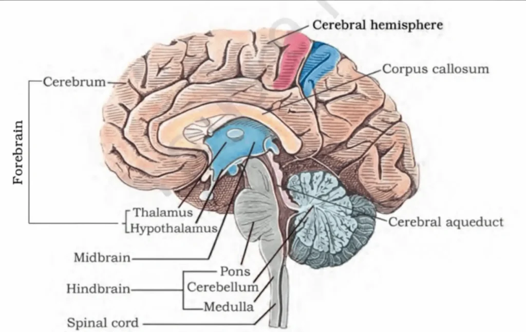
| Part of Brain | sub-parts | Structure & Functions |
| Fore Brain | Cerebrum | Most developed & largest part of forebrain (about 2/3 of brain)Divided into Right & Left parts = Cerebral hemispheresConnected by Corpus callosum(Outer surface folded with ridges & groovesFunctions: Thinking, reasoning, planning, memorizing, initiates contraction of voluntary muscles & controls them |
| Olfactory lobes | Two lobes at anterior side of cerebral hemisphereHelp in sense of smell | |
| Diencephalon | Extremely sensitiveThalamus: Transmits sensory impulses (pain, comfort) to cerebrum.It is the centre of the pain, cold and heat.Hypothalamus: It controls the hormonal secretion of endocrine glands. Hormones secreted from posterior pituitary gland secrach through it. This is the centre of hunger, thirst, temperature control, love, hate etc. Blood pressure, metabolism of water, sweat, anger, joy etc are controlled by it. | |
| Mid Brain | Corpora Quadrigemina | Two superior optic lobes + Two inferior lobesReceive stimuli related with vision & hearing |
| Cerebral peduncle | Structural part of midbrain | |
| HindBrain | Cerebellum | Maintains body balanceMaintains coordination in muscular activities |
| Pons Varolii | Contains pneumotaxic center → regulates respiratory ventilationMediator controller in chewing, salivation, hearing, secretion of tears, movement of eyeballs | |
| Medulla Oblongata | Last part of brain, attached to spinal cordFunctions: Breathing, coughing, swallowing, heartbeats, blood pressure, peristaltic movement of alimentary canal & other involuntary activities |
- Note : EEG (Electroencephalography) is done to known the function of brain.
- Spinal cord : The posterior region of the medulla oblongata forms the spinal cord. Its main functions are :
- (a) Coordination and control of reflex actions i.e. it works as the centre of the reflex actions.
- (b) It carries the impulses coming out of brain.
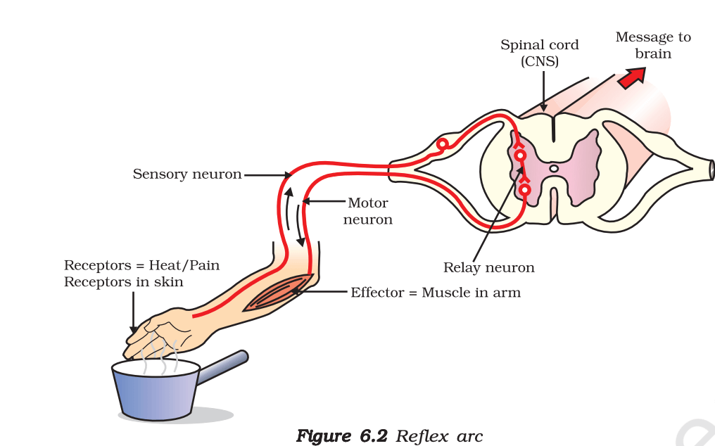
Note : Reflex action was first discovered by the scientist, Marshall Hall.
Reflex action
- Reflex Arch – pathway involved
- The Central Nervous system consists of the Brain and spinal cord, Brain does not take part in immediate reflex action.
- The spinal cord, which reacts without thinking about how to respond to stimuli, is in charge of these automatic actions
- Pathway involved
- Receptor– It receives the information from Stimuli.
- Sensory Nerve It carries information from receptor to spinal cord,
- Spinal cord(Interneurons) It transfers the information from sensory nerve to the Motor nerve. It is present in the spinal cord.
- Motor nerve It carries the information from the spinal cord to effective organs (muscles).
- Effector muscles. It receives the information from Motor Nerve & show their effect
- Exa: Response to any hot object, response to needle, saliva secretion by seeing delicious food.
Peripheral Nervous System :
- Peripheral Nervous System is made up of the nerves arising from brain and spinal cord. These are called cranial and spinal nerves respectively. There are sensory, motor and mixed nerve.
- There are 12 pairs of cranial nerves and 31 pairs of spinal nerve found in a human.
- The unit of nervous tissues is called Neuron or nerve cell.
Autonomic Nervous System
- Autonomic Nervous System is made up of some brain nerves and some spinal cord nerves. It supplies nerves to all the internal organs and blood vessel of the body. There are two parts of Autonomic Nervous System :
(a) Sympathetic Nervous System
Functions
- It reduces the secretion of salivary glands.
- It increases the heart beat. It increases the secretion of sweat glands.
- The rate of respiration increase. It increases the blood pressure.
- It increases the sugar level in the blood.
- Collective impact of this affects fear, pain and anger.
(b) Parasympathetic Nervous System
Functions
- The functions of this system is normally the opposite of Sympathetic Nervous System.
- For example : It increases the secretion of saliva and other digestive juices. The effect of this nervous system collectively creates the occasion of rest and joy.
Skeletal System
- The skeletal system of human is made up of two parts : (a) Axial skeleton (b) Appendicular skeleton.
Axial skeleton :
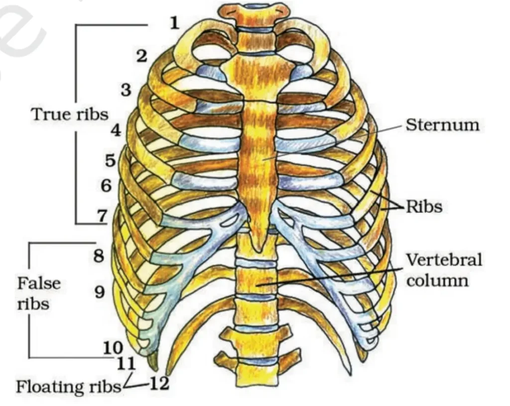
- The skeleton, which makes the main axis of the body is called axial skeleton. Skull, vertebral column and bones of chest comes under it. There are 80 bones in axial skeleton.
- Skull : There are 29 bones in it. Out of these, 8 bones jointly protect the brain of the human. The structure made up of these bones is called forehead. All the bones of the forehead remain joined strongly by the sutures. There are 14 bones in addition to this which form the face. Six ear ossicles of both the ears and one hyoid bone.
- Vertebral Column : The vertebral column of the human is made up of 33 vertebra. All the vertebra are joined by intervertebral disc. Vertebra is made flexible by these intervertebral disc. Its first vertebra which is called atlas holds the skull.
Appendicular skeleton :
- The following are the parts of it—
- Foot bones— Both hands and feet have 118 bones.
- To hold the forelimb and hind limb on the axial skeleton in human there are two girdles
- The girdle of forelimb is called pectoral girdle and girdle of hindlimb is called pelvic girdle.
- Pectoral girdle joined with forelimb is called humerus and the bone from pelvic girdle join to hindlimb is called femur.
Note:-
- The total number of bones in a human body – 206
- The total number of bones during childhood – 300
- The total number of bones of head – 29 (forehead–8, facial–14, ear–6, hyoid–1)
- The total number of bones in vertebral column, initially–33, After development – 26 (5 sacral fuse into 1 and 4 caudal fuse into 1)
- The total number of bones of ribs – 24
- The longest and strongest bone of the body – Femur (bone of thigh)
- The smallest bone of the body – Stapes (bone of ear)
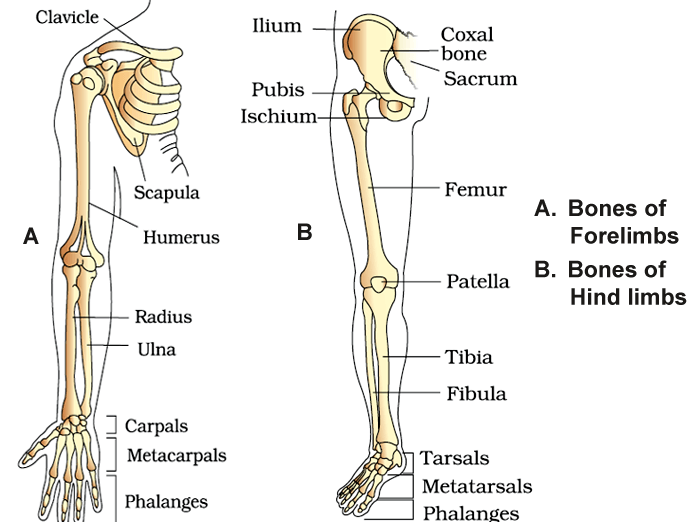
- Arm bones ( 30 )- Humerus ,Radius, ulna, 8 carpals in wrist of hand ,5 metacarpals bones located in palm,14 Phalanges in fingers 2 in thumb 3 in each finger.
- Leg bones ( 30 ) – Femur, Tibia and fibula ,7 tarsals, Five Metatarsals, 14 Phalanges ,Triangular and flat sesamoid bone of knee is patella
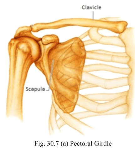
- Pectoral Girdle (Shoulder Girdle)
- It connects the arms (upper limbs) to the body.
- It has two separate halves (right and left).
- Each half has two bones: Clavicle (collar bone), Scapula (shoulder blade)
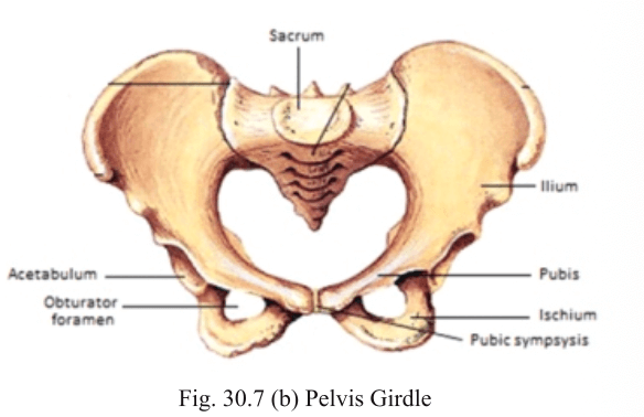
- Pelvic Girdle (Hip Girdle)
- It connects the legs (lower limbs) to the body.
- It has two halves but both are joined in the middle at pubic symphysis.
JOINTS
- Joints are essential for all types of movements involving the bony parts of the body.
Classification of Joints
- Fibrous Joints – Do not allow movement. Example: Skull bones joined by sutures.
- Cartilaginous Joints – Bones are joined by cartilage.Allow limited movement. Example: Joints between vertebrae.
- Synovial Joints – Have a fluid-filled cavity between bones. Allow free movement. Examples: Ball and socket – shoulder, Hinge – knee, Pivot – atlas and axis, Gliding – wrist, Saddle – thumb
Major Types of Joints in the Human Body
| Type of Joint | Example | Bones Involved |
| Ball and Socket Joint | Shoulder, Hip | Shoulder: Humerus & Pectoral girdle (Scapula) Hip: Femur & Pelvic girdle |
| Hinge Joint | Elbow, Knee, Ankle, Fingers | Elbow: Humerus & Ulna Knee: Femur & Tibia Ankle: Tibia & Talus |
| Pivot Joint | Neck (Atlas & Axis), Radioulnar joint | Atlas & Axis vertebrae Radius & Ulna |
| Saddle Joint | Thumb (Carpometacarpal joint of thumb) | Carpal (Trapezium) & First Metacarpal (Thumb) |
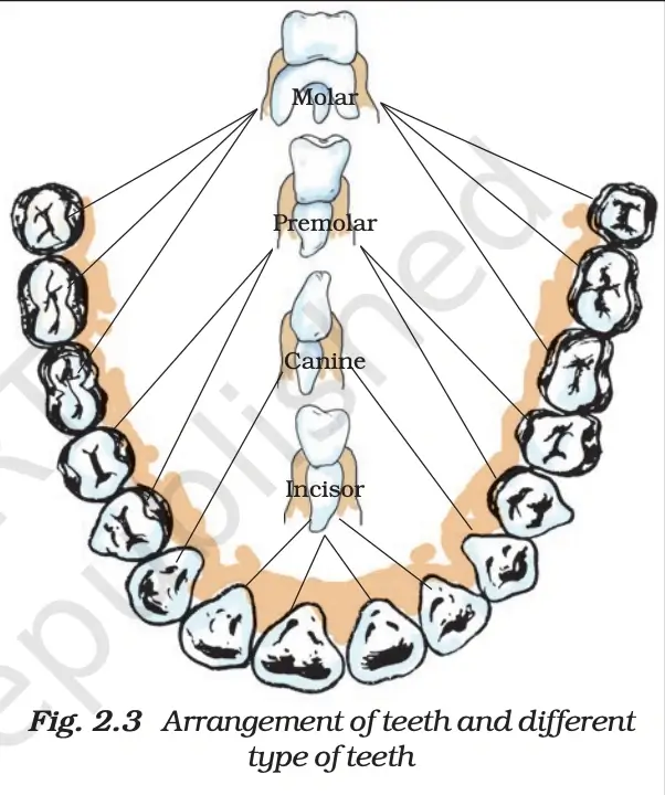
Tooth
- Types of teeth – Incisors ,Canion, premolar ,Molar
- Dental formula = teeth in upper jaw/ teeth in lower jaw × 2
- Dental formula – 2123/2123 × 2 (32 teeth)
- Before wisdom teeth – 2122/2122 × 2 (28 teeth)
- The number of teeth in the upper jaw of a 13 year old child – 14
- Milk’s teeth – 2102/2102 × 2 (20 teeth)
- 2nd incisor teeth of elephant grow and became tusk
DISORDERS OF MUSCULAR AND SKELETAL SYSTEM
- Myasthenia gravis: Auto immune disorder affecting neuromuscular junction leading to fatigue, weakening and paralysis of skeletal muscle.
- Muscular dystrophy: Progressive degeneration of skeletal muscle mostly due to genetic disorder.
- Tetany: Rapid spasms (wild contractions) in muscle due to low Ca++ in body fluid.
- Arthritis: Inflammation of joints.
- Osteoporosis: Age-related disorder characterised by decreased bone mass and increased chances of fractures. Decreased levels of estrogen is a common cause
- Gout: Inflammation of joints due to accumulation of uric acid crystals.
Circulatory System
- The discovery of blood circulation was done by William Harvey in 1628.
- There are four parts under it – (a) Heart (b) Arteries (c) Veins (d) Blood
- Heart
- It remains safe in the pericardial membrane. Its weight is approximately 300 grams.
- Heart of the human is made up of four chambers. In the anterior side there is a right auricle and a left auricle. In the posterior side of the heart a right ventricle and a left ventricle persist.
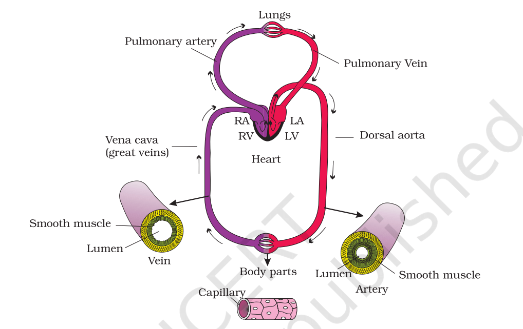
- Between the right auricle and the right ventricle there is a tricuspid valve.
- Between the left auricle and left ventricle there is a bicuspid valve.
Course of circulation
- Mammals have double circulation. It mean blood have to cross two times from heart before circulating throughout the body.
- Right auricle receive impure blood from the body which goes into right ventricle. From here the blood goes into pulmonary artery which send it to the lungs for purification. After purification it is collected by pulmonary vein which brings it back to heart in left auricle. From auricle it goes into left ventricle. Now this purified blood is carried by arota to different organ of body. This circulation is done is a cardiac cycle.
Double Circulation
- In humans, blood passes two times through the heart in one full round. This is called double circulation.
- (A) Pulmonary Circulation
- Deoxygenated blood → Right ventricle → Pulmonary artery
- Blood goes to lungs, Blood becomes oxygenated
- Oxygenated blood → Pulmonary veins → Left atrium
- (B) Systemic Circulation
- Oxygenated blood → Left ventricle → Aorta
- Blood goes to all body parts
- Oxygen and nutrients are supplied
- Deoxygenated blood comes back to right atrium through veins
Cardiac Cycle
- The cardiac cycle means all the activities that take place in the heart during one heartbeat.
- Time for one cardiac cycle = 0.8 seconds
- Blood pumped by one ventricle in one beat = 70 mL (Stroke Volume)
- Heart Sounds:
- Lub → Sound of AV valves closing
- Dub → Sound of semilunar valves closing
Electrocardiogram (ECG)
- ECG is a graph that shows the electrical activity of the heart.
- It is recorded using a machine called electrocardiograph.
- ECG helps doctors check if the heart is working normally.
Terms
- Vein – This blood vessel carries the blood from the body towards the heart is called vein. In vein there is impure blood i.e. carbon dioxide mixed blood. Its exception is the pulmonary vein, which always carries oxygenated blood.
- Artery – These blood vessels carry blood from the heart to other parts of the body .In the artery there is oxygenated blood. Its exception is pulmonary artery.
- Sinoatrial node (SA node) : It is a specialised area of cardiac muscle fibre in right auricle. SA node is also known as pacemaker as it generates each wave of cardiac impulse.
- Atrioventricular node (AV node) : AV node is present close to the interatrial septum near the right AV aperture.
- Artificial pacemaker : When SA node becomes defective or damaged, the cardiac impulses do not generate. This can be cured by surgical grafting of an artificial pacemaker an electric device in the chest of the patient. It stimulate the heart electrically at regular intervals.
- Systole and diastole of the heart are collectively called heart beat. In the normal condition the heart of the human beats 72 times and in a single beat it pumps approximately 70 ml blood.
- The blood pressure of a normal human is 120/80. (Systolic – 120 and Diastolic – 80).
- Blood pressure is measured by sphygmomanometer.
- Thyroxin and adrenaline are the hormones which independently controls the heart beat.
Disorders of Circulatory System
- High Blood Pressure (Hypertension)
- Normal BP = 120/80 mm Hg
- Hypertension = 140/90 mm Hg or more
- It can damage: Heart, Brain, Kidneys
- Coronary Artery Disease (CAD)
- Fat, cholesterol, calcium deposit in arteries.
- Blood supply to heart muscles is reduced.
- Can cause heart attack.
- Angina Pectoris
- Sudden chest pain.
- Due to lack of oxygen to heart muscles.
- Common in older people.
- Heart Failure
- Heart cannot pump enough blood
- Lungs get filled with fluid.
- It is different from heart attack and cardiac arrest.
