
Magnetic Resonance Imaging (MRI) is an advanced medical imaging technique explained in Physics that uses strong magnetic fields and radio waves to produce detailed images of internal body structures. It is widely used in healthcare because it is non-invasive and provides clear, high-resolution pictures of organs and tissues.
Previous year Questions
| Year | Question | Marks |
| 2023 | Discuss in brief the principle and working of MRI. Why it is considered safer thanx-ray tomography ? | 10M |
Magnetic Resonance Imaging (MRI)
Magnetic resonance imaging (MRI) is a non-invasive medical imaging technique that uses a magnetic field and radio pulses to generate detailed images of the internal structures of the body.MRI utilizes proton NMR to image the concentration of protons, making it ideal for imaging soft tissues like the brain and eyes. Tissues with high proton density appear brighter in the images, while those with low proton density, such as bone, appear dark.
MRI Enhancements Over NMR
- Spatial Localization:
- NMR provides molecular data for small samples, while MRI introduces gradient magnetic fields to spatially localize signals and create images.
- Image Construction:
- MRI does not just detect the signals, but also reconstructs cross-sectional or 3D images of tissues, allowing detailed visualization of internal structures.
- Clinical Relevance:
- MRI offers detailed soft tissue imaging that is crucial in diagnosing diseases, injuries, and conditions in humans.
Basic Principles of MRI
- Nuclear Spins in MRI:
- The nucleus of hydrogen atoms in the body (mostly found in water and fat) has a magnetic moment due to its intrinsic spin.
- In an external magnetic field (B0B0), these hydrogen nuclei align either parallel or anti-parallel to the field, which creates energy states.
- The energy difference between these states is small, and the magnetic field causes the spins to precess (rotate) around the axis of the field.
- Larmor Frequency:
- Similar to NMR, hydrogen nuclei precess at a frequency called the Larmor frequency, which is proportional to the strength of the magnetic field.
- The Larmor frequency (ω0) is given by:
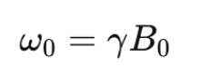
- γ is the gyromagnetic ratio for hydrogen (which is constant), and B0 is the strength of the magnetic field.
Key Components of an MRI System
A. Magnetic Field:
- The primary component of an MRI machine is a superconducting magnet, which creates a powerful magnetic field.
- Strength: MRI machines typically use magnetic fields of 1.5 Tesla to 3 Tesla, but research machines can go up to 7 Tesla or higher.
- Function: The magnetic field aligns the hydrogen nuclei (protons) in the human body, especially those in water and fat molecules.
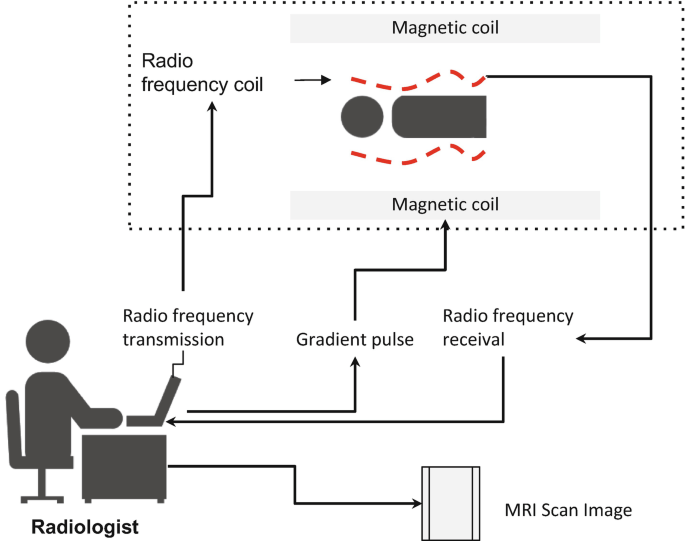
B. Radiofrequency (RF) Pulses:
- Purpose: RF pulses are used to excite the aligned hydrogen nuclei, causing them to absorb energy and flip to a higher energy state.
- How it Works:
- The magnetic field causes the hydrogen protons to align either parallel or anti-parallel to the magnetic field.
- A short RF pulse is applied at a specific frequency to knock the protons out of alignment.
- When the RF pulse is turned off, the protons return to their original alignment, emitting energy in the form of RF signals.
C. Gradient Coils:
- Magnetic field gradients are applied in three directions: X-axis, Y-axis, and Z-axis.
- Function: These gradients vary the magnetic field strength in different regions of the body, allowing MRI to determine the location of the signal from different parts of the body.
- Purpose: They help localize signals in space by encoding positional information, allowing the machine to generate 2D or 3D images.
D. Receiver Coil:
- Relaxation: After the RF pulse is turned off, the hydrogen nuclei return to their equilibrium state (lower energy state) by releasing energy, which is detected as an NMR signal. This process of returning to equilibrium is known as relaxation.
- Function: Receiver coil detects these RF signals emitted by the hydrogen protons during relaxation.
E. Image Reconstruction:
- The collected signals are sent to a computer that processes the data and reconstructs the image.
- This is done through a process called Fourier Transform, which converts the frequency and phase data into spatial information (creating the image).
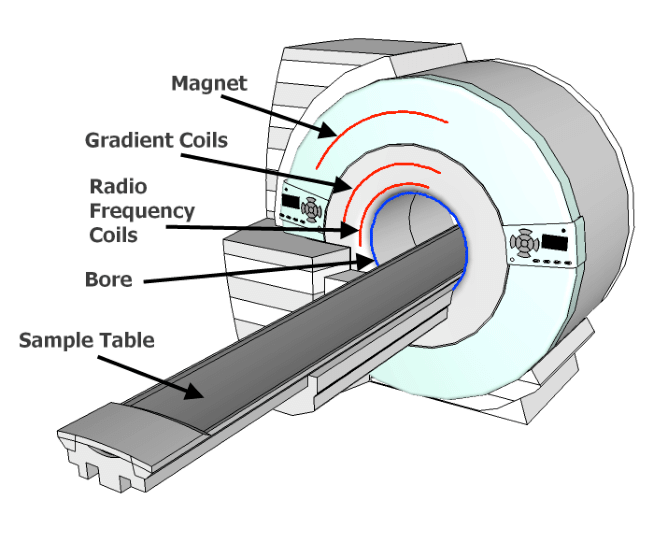
How MRI Works
- Magnet → Strong magnetic field aligns hydrogen protons in the body.
- RF Coil → RF pulse flips protons out of alignment.
- Gradient Coils → Varying magnetic fields map the location of protons.
- Receiver Coil → Protons relax and emit RF signals.
- Computer → Signals are processed to create detailed images.
Types of MRI Scans
- T1-weighted Imaging:
- Useful for anatomical imaging where fat and other solid tissues appear bright.
- Primarily used for structural imaging, such as brain scans and joint examinations.
- T2-weighted Imaging:
- Excellent for imaging areas with high water content, such as edema or inflammation.
- T2-weighted images highlight fluid-filled spaces and are particularly useful for detecting pathology such as tumors and infections.
- Functional MRI (fMRI):
- Measures brain activity by detecting changes in blood oxygenation (BOLD – Blood Oxygen Level Dependent contrast).
- Used for mapping brain functions and studying neural activity in response to various stimuli.
- Diffusion-Weighted Imaging (DWI):
- Measures the movement of water molecules in tissues, commonly used for detecting strokes or tumors.
Applications of MRI
1. Medical Diagnostics:
- Brain Imaging: MRI is widely used for diagnosing brain conditions such as tumors, strokes, and neurological disorders(e.g., multiple sclerosis).
- Musculoskeletal Imaging: MRI is crucial for evaluating injuries to muscles, ligaments, tendons, and joints (e.g., ACL tears, fractures, arthritis).
- Cardiac Imaging: Cardiac MRI is used to assess heart conditions, such as heart failure, congenital heart defects, and myocardial infarction.
- Spinal Cord Imaging: Used to diagnose spinal cord injuries and disorders like herniated discs, spinal tumors, and degenerative diseases.
- Oncology: MRI helps in detecting cancers, particularly those affecting soft tissues. It helps in locating tumors and assessing their size and spread.
- Abdominal Imaging: MRI helps in visualizing internal organs like the liver, kidneys, pancreas, and bowels, often for detecting tumors, cysts, or infections.
2. Functional MRI (fMRI):
- Brain Activity Mapping:
- fMRI measures brain activity by detecting changes in blood oxygenation levels. It is used to map regions of the brain involved in specific tasks (e.g., motor movement, speech, vision).
- It plays a crucial role in understanding neuroplasticity and brain function, particularly in pre-surgical planning for brain tumors or epilepsy.
Advantages of MRI
- Non-invasive: No need for surgery to view internal organs and tissues.
- Non-radiative: Unlike X-rays or CT scans, MRI does not use ionizing radiation, making it safer for repeated use, especially in younger patients.
- High Soft Tissue Contrast: MRI excels at imaging soft tissues like the brain, spinal cord, muscles, and organs. It provides superior contrast for these tissues compared to X-rays or CT scans.
- Versatility: MRI can be adapted to image different types of tissues and conditions using various specialized techniques (e.g., contrast-enhanced MRI, functional MRI, diffusion tensor imaging).
- No Need for Contrast in Some Cases: MRI scans are capable of providing high-quality images of soft tissues without the need for contrast agents in some instances (e.g., in brain imaging).
Limitations of MRI
- Cost and Accessibility:
- MRI machines are expensive to purchase, maintain, and operate, making them less accessible in low-resource settings.
- Time-Consuming:
- MRI scans take a relatively long time compared to other imaging techniques (e.g., CT scans), typically ranging from 15 minutes to an hour.
- Contraindications for Certain Patients:
- MRI is not suitable for patients with metal implants, pacemakers, or other magnetic-sensitive devices.
- The strong magnetic field can interfere with implanted devices and pose safety risks.
- Sensitivity to Movement:
- Patients must remain very still during the scan. Movement can blur the images, leading to inaccurate results.
- Limited for Bone Imaging:
- MRI is not as effective as CT scans for visualizing bone fractures or other bone-related conditions, as it provides superior detail for soft tissues.
- Claustrophobia:
- Some patients feel uncomfortable or claustrophobic inside the narrow MRI machine, which can make it difficult for them to stay still during the scan.
MRI Safety
- Magnetic Field: The powerful magnets in MRI machines can interfere with metal objects and medical implants. Patients are thoroughly screened before undergoing an MRI.
- Contrast Agents: Some MRI scans require a contrast agent (usually gadolinium-based) to help highlight certain areas. Though generally safe, these agents can cause allergic reactions in rare cases.
Differences Between NMR and MRI
| Aspect | NMR (Nuclear Magnetic Resonance) | MRI (Magnetic Resonance Imaging) |
| Purpose | Study molecular and atomic structures | Non-invasive medical imaging of tissues and organs |
| Sample Type | Small chemical samples (liquids or solids) | Human body or animal tissues |
| Signal Detected | Chemical shifts and molecular interactions | Proton density and relaxation times (T1, T2) of tissues |
| Resolution | Provides high-resolution spectra for molecular analysis | Provides high-resolution 2D or 3D images of internal organs |
| Main Application | Chemistry, biochemistry, material science | Medicine (diagnostic imaging) |
FAQ (Previous year questions)
Principle of MRI (Magnetic Resonance Imaging):
MRI is based on Nuclear Magnetic Resonance (NMR). When placed in a strong magnetic field, hydrogen nuclei (protons) in the body align with the field. These nuclei absorb and emit radiofrequency (RF) energy when disturbed by an RF pulse. The emitted signals are detected and used to construct images.
Working of MRI:
Component
Function
Magnet
Aligns hydrogen protons in the body using a strong magnetic field (1.5–3 T).
RF Coil
Sends pulses to excite protons, flipping them from alignment.
Gradient Coils
Vary magnetic fields to localize the position of protons.
Receiver Coil
Detects signals emitted as protons relax to original alignment.
Computer System
Converts signals into detailed 2D/3D images using Fourier Transform.
Why MRI is Safer than X-ray Tomography:
Parameter
MRI
X-ray/CT Scan
Radiation
No ionizing radiation
Uses ionizing radiation (X-rays)
Soft Tissue Imaging
High contrast for soft tissues
Lower soft tissue contrast
Safety for Repeated Use
Safe for multiple scans
Cumulative radiation exposure risk
Biological Risk
Minimal (non-ionizing RF waves)
Potential DNA damage from ionizing radiation
MRI is a non-invasive, radiation-free imaging technique with high soft tissue contrast, making it ideal for neurological, musculoskeletal, and organ imaging. Its superiority in safety and resolution, particularly in repeated diagnost
