
Circulatory and Digestive Systems in Human Being are two vital biological processes that keep our body functioning efficiently. In Biology, the circulatory system transports oxygen, nutrients, and hormones throughout the body, while the digestive system breaks down food into simpler forms for energy and growth. Together, they ensure proper nourishment and overall health.
Circulatory system
Types of Circulatory Systems:
The circulatory system of animals can be broadly categorized into two types:
- Open Circulatory System:
- Present in: Arthropods (e.g., insects, spiders) and mollusks (e.g., snails, clams).
- Mechanism: In an open circulatory system, the blood (called hemolymph) is pumped by the heart through large vessels into open spaces or body cavities, known as sinuses. These sinuses surround the organs and tissues. The hemolymph directly bathes the cells, allowing for nutrient and gas exchange.
- Advantage: Simpler and less energy-intensive, but less efficient in regulating blood flow.
- Closed Circulatory System:
- Present in: Annelids (e.g., earthworms), chordates (including vertebrates like fishes, amphibians, reptiles, birds, and mammals).
- Mechanism: In a closed circulatory system, blood is pumped by the heart through a network of closed blood vessels. The blood never leaves the vessels, and oxygen, nutrients, and wastes are exchanged through the vessel walls to the surrounding tissues.
- Advantage: More efficient, as the flow of blood can be precisely regulated, leading to better control of oxygen and nutrient delivery to tissues.
Human Circulatory System :
The human circulatory system moves blood around the body and consists of the heart, blood vessels, and blood.
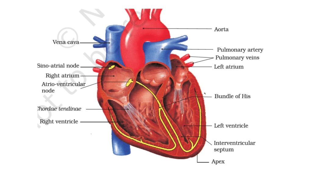
Heart Structure
- The heart is located in the thoracic cavity, between the two lungs, slightly tilted to the left.
- The heart is protected by a double-walled membranous bag called the pericardium, which contains pericardial fluid that reduces friction.
- Four chambers make up the heart:
- Two atria (upper chambers): Smaller and thinner-walled.
- Two ventricles (lower chambers): Larger and thicker-walled.
Septae and Valves
- Septae are walls that separate the heart chambers.
- Valves ensure blood moves in one direction, preventing any backward flow:
- Tricuspid valve: Between the right atrium and right ventricle.
- Bicuspid (Mitral) valve: Between the left atrium and left ventricle.
- Semilunar valves: Prevent blood from flowing backward into the ventricles.
Cardiac Muscle and Nodal Tissue
- The heart is made of cardiac muscle. Specialized muscle tissue called nodal tissue is responsible for initiating the heart’s rhythmic contractions:
- Sino-atrial node (SAN): The heart’s natural pacemaker, it starts the heartbeat.
- Atrioventricular node (AVN): Passes the signal to the ventricles, causing them to contract.
- The heart beats around 70-75 times per minute to keep blood flowing.
Human heart
Cardiac Cycle
The cardiac cycle is the series of events that happen in one heartbeat, involving contraction (systole) and relaxation (diastole) of the heart chambers.
- Joint Diastole: All chambers are relaxed. Blood flows into the atria from veins (vena cava on the right and pulmonary veins on the left).
- Atrial Systole: The atria contract (triggered by the SAN), pushing blood into the ventricles.
- Ventricular Systole: The ventricles contract (stimulated by the AVN), closing the tricuspid and bicuspid valves and opening the semilunar valves. Blood is pumped into the pulmonary artery (right side) and aorta (left side).
- Ventricular Diastole: The ventricles relax, the semilunar valves close, and blood refills the atria and ventricles.
A full cardiac cycle lasts 0.8 seconds and includes both atrial and ventricular systole and diastole.
- Stroke volume: The amount of blood pumped by each ventricle per beat (~70 mL).
- Cardiac output: The volume of blood pumped by each ventricle per minute (calculated as stroke volume × heart rate). For a healthy adult, it averages 5 liters per minute.
Heart Sounds:
- The first heart sound (Lub):: When valves between atria and ventricles close. {closure of the tricuspid and bicuspid valves}
- Dub: When valves leading to arteries close. {closure of the semilunar valves
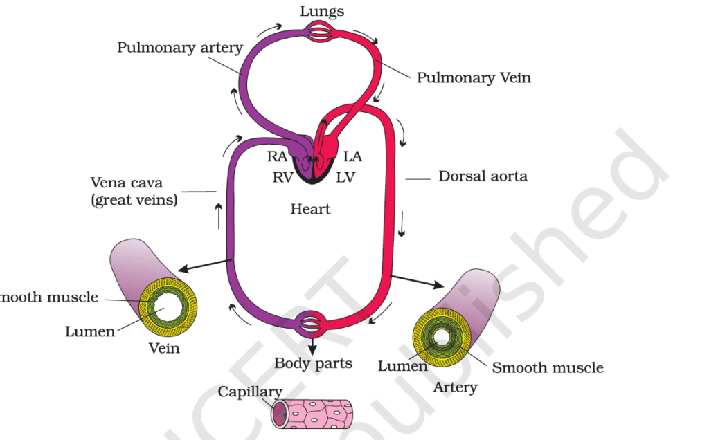
Blood Vessels:
Blood vessels are pathways for blood circulation. There are three main types:
- Arteries:
- Structure: Thick, muscular, and elastic walls to handle high pressure.
- Function: Carry oxygen-rich blood from the heart to the body (except the pulmonary artery, which carries oxygen-poor blood to the lungs).
- Key Arteries:
- Aorta: Largest artery, supplies oxygenated blood to the body.
- Pulmonary artery: Carries deoxygenated blood to the lungs.
- Veins:
- Structure: Thin walls with valves to prevent backflow of blood.
- Function: Carry oxygen-poor blood back to the heart (except pulmonary veins, which bring oxygen-rich blood from the lungs).
- Key Veins:
- Vena cava: Largest veins, bring deoxygenated blood to the heart.
- Pulmonary veins: Carry oxygenated blood to the heart.
- Capillaries:
- Structure: Extremely thin, one-cell-thick walls.
- Function: Allow exchange of oxygen, nutrients, and waste between blood and tissues.
- Capillary Networks: Dense networks ensure all cells get oxygen and nutrients.
These vessels work together to transport blood efficiently throughout the body.
Double Circulation:
In humans, blood flows through the heart twice during one complete circuit. This ensures efficient oxygen delivery and waste removal.
- Pulmonary Circulation:
- Deoxygenated blood is pumped from the right ventricle to the lungs through the pulmonary artery.
- In the lungs, blood picks up oxygen and releases carbon dioxide.
- Oxygenated blood returns to the left atrium via the pulmonary veins.
- Systemic Circulation:
- Oxygenated blood moves from the left atrium to the left ventricle and is pumped into the aorta.
- Arteries, arterioles, and capillaries deliver oxygen to tissues and collect carbon dioxide.
- Deoxygenated blood returns to the right atrium through veins.
This dual system ensures continuous oxygen supply to the body and removal of waste gases.
Pulmonary Circulation:
- Right Ventricle → Pulmonary Artery → Lungs (Oxygen in, CO₂ out)
- Pulmonary Veins → Left Atrium
Systemic Circulation:
- Left Atrium → Left Ventricle → Aorta → Arteries → Capillaries (Oxygen to tissues, CO₂ to blood)
- Veins → Right Atrium
Special Circulatory Pathways:
- Hepatic Portal System:
- This system carries blood from the digestive organs to the liver before it enters the general circulation. It ensures that nutrients absorbed from the digestive tract are processed by the liver before being distributed to the rest of the body.
- Coronary Circulation:
- The coronary arteries supply the heart muscle with oxygen-rich blood. Blood from the heart muscle returns to the right atrium via the coronary veins.
- Blockage or narrowing of these arteries can lead to heart conditions like angina or heart attacks (myocardial infarction).
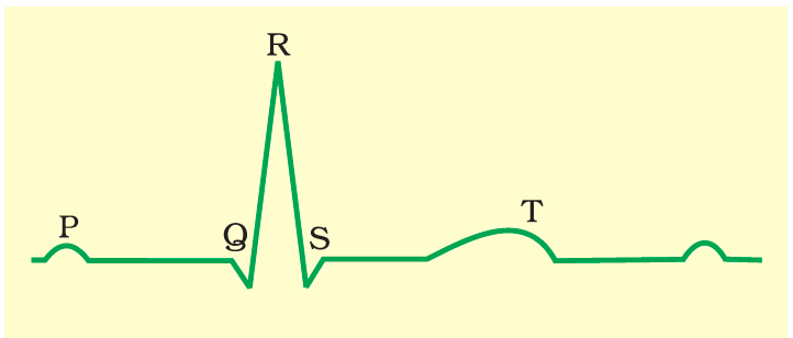
Electrocardiogram (ECG)
An ECG is a graphical representation of the heart’s electrical activity during the cardiac cycle. It is used to assess heart function.
- P-wave: Represents atrial depolarization (atrial contraction).
- QRS complex: Represents ventricular depolarization (ventricular contraction).
- T-wave: Represents ventricular repolarization (ventricular relaxation)
Regulation of Cardiac Activity
Intrinsic Regulation (Myogenic Regulation):
- The heart has its own built-in rhythm, generated by specialized muscle tissue known as nodal tissue:
- Sinoatrial (SA) Node: Located in the right atrium, it sets the heart’s rhythm and is called the heart’s “pacemaker.”
- Atrioventricular (AV) Node: Located between the atria and ventricles, it delays the signal slightly to ensure the atria fully contract before the ventricles.
- Bundle of His & Purkinje Fibers: These transmit the electrical signal to the ventricles, causing them to contract.
Extrinsic Regulation (Neural and Hormonal Regulation):
- Autonomic Nervous System (ANS): The ANS adjusts heart function with two branches:
- Sympathetic Nervous System: Increases heart rate and contraction strength during stress or physical activity (the “fight or flight” response).
- Parasympathetic Nervous System: Slows down the heart rate and reduces contraction strength, especially during rest.
Hormonal Regulation:
- Adrenaline (and noradrenaline) increase heart rate and force of contraction during stress.
- Thyroid hormones can make the heart more sensitive to sympathetic signals, increasing heart rate.
Disorders of the Circulatory System:
1. Hypertension (High Blood Pressure)
- Definition: Consistently high blood pressure (above 140/90 mm Hg).
- The systolic pressure (120 mm Hg) represents the pressure when the heart contracts, and the diastolic pressure(80 mm Hg) reflects the pressure when the heart is at rest.
- Causes: Excess salt, obesity, stress, genetics, aging.
- Consequences: Damages heart and blood vessels, leading to heart disease, strokes, kidney problems, and eye issues. It can cause heart failure and coronary artery disease over time.
2. Coronary Artery Disease (CAD)
- Definition: Narrowing or blockage of the coronary arteries due to fatty deposits (plaques).
- Causes: Buildup of fat, cholesterol, and other substances in the arteries.
- Consequences: Reduced blood flow to the heart, leading to chest pain (angina) and potentially heart attacks.
3. Angina (Angina Pectoris)
- Definition: Chest pain caused by reduced oxygen to the heart muscle.
- Types:
- Stable Angina: Predictable and subsides with rest.
- Unstable Angina: Unpredictable, can occur at rest, and may signal an impending heart attack.
- Cause: Often due to narrowed coronary arteries from atherosclerosis.
4. Heart Failure
- Definition: When the heart cannot pump blood efficiently, leading to fluid buildup in the lungs, abdomen, and legs.
- Causes: Coronary artery disease, hypertension, heart attacks, valvular heart disease.
- Symptoms: Shortness of breath, fatigue, swelling, fluid retention.
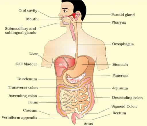
Digestive System
Digestive System: Alimentary Canal + Digestive Gland
Alimentary canal
The alimentary canal is a continuous tube in the digestive system that begins with the mouth (anterior opening) and ends at the anus (posterior opening).
Mouth and Buccal Cavity
- The mouth opens into the buccal cavity (oral cavity), which plays a crucial role in mechanical and chemical digestion.
- The oral cavity contains:
- Teeth: Specialized for chewing and breaking down food into smaller particles.
- Muscular tongue: Helps manipulate food and is essential for swallowing and speech.
Teeth
- Thecodont Attachment:
- Each tooth is embedded in a socket within the jawbone. This type of attachment is known as the codont.
- Diphyodont Dentition:
- Humans and most mammals form two sets of teeth during their lifetime:
- Deciduous Teeth (milk teeth): Temporary teeth that erupt in early childhood.
- Permanent Teeth: Replace the milk teeth and remain for life, unless lost due to injury, disease, or aging.
- Humans and most mammals form two sets of teeth during their lifetime:
Total Teeth: Adult humans have 32 permanent teeth.
Types of Teeth (Heterodont Dentition):
- Incisors (I): For cutting food.
- Canines (C): For tearing food.
- Premolar (PM) and Molars (M): For grinding and crushing food.
Dental Formula: Represents the arrangement of teeth in one half of each jaw (upper and lower).
Enamel: Hard, outer chewing surface of teeth, essential for mastication (chewing).
Tongue
- Structure:
- Freely movable muscular organ.
- Attached to the floor of the oral cavity by the frenulum.
- Upper surface contains papillae, some of which have taste buds.
- Functions:
- Manipulates food for chewing and swallowing.
- Houses taste buds for detecting flavors
Pharynx
- A short common passage for both food and air.
- Connects the oral cavity to the oesophagus and trachea.
- Epiglottis: A cartilaginous flap that prevents food from entering the trachea during swallowing.
Oesophagus
- A thin, long muscular tube extending from the pharynx to the stomach.
- Passes through the neck, thorax, and diaphragm.
- The gastro-oesophageal sphincter regulates the opening of the oesophagus into the stomach.
Stomach
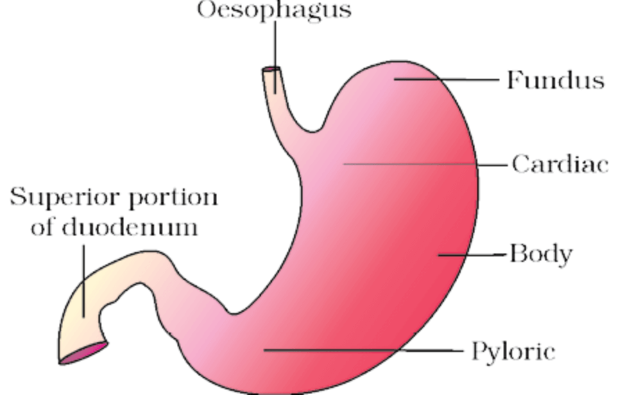
- A J-shaped organ located in the upper left abdominal cavity.
- Parts of the Stomach:
- Cardiac Portion: Where the oesophagus opens.
- Fundic Region: Dome-shaped upper part.
- Body: Main central region.
- Pyloric Portion: Opens into the small intestine via the pyloric sphincter.
Small Intestine
- Divided into three regions:
- Duodenum: C-shaped, first section.
- Jejunum: Long, coiled middle section.
- Ileum: Highly coiled distal section, opens into the large intestine.
Large Intestine
- Consists of:
- Caecum:
- A small, blind sac.
- Hosts symbiotic micro-organisms.
- Includes the vermiform appendix (a vestigial organ).
- Colon:
- Divided into ascending, transverse, descending, and sigmoid parts.
- Rectum:
- Stores feces temporarily.
- Opens out through the anus.
- Caecum:
Digestive Glands:
The digestive glands play a vital role in producing secretions that aid in the digestion of food.
Salivary Glands
- Types:
- Parotids: Located near the cheeks.
- Sub-maxillary/Sub-mandibular: Located in the lower jaw.
- Sub-linguals: Located below the tongue.
- Function: Secrete salivary juice into the buccal cavity.
Composition of Saliva:
- Water (99.5%): Moistens food and dissolves it for taste perception.
- Mucus: Lubricates the bolus (chewed food) to facilitate swallowing.
- Enzymes: → Salivary Amylase (Ptyalin):
- Breaks down starch into maltose and dextrins.
- Begins carbohydrate digestion in the mouth.
- Lysozyme: An antimicrobial enzyme that protects against bacteria.
Function of Saliva:
- Initiates the digestion of starch.
- Facilitates mastication and swallowing.
- Keeps the oral cavity moist and prevents microbial growth.
Liver
- Structure: Largest gland in the body, weighing 1.2–1.5 kg in adults.
- Located in the abdominal cavity just below the diaphragm.
- Composed of two lobes.
- Hepatic Lobules:
- Functional and structural units of the liver.
- Contain hepatic cells arranged in cords.
- Bile Production:
- Secreted by hepatic cells and transported via hepatic ducts.
- Stored and concentrated in the gall bladder.
- Functions:
- Bile facilitates the emulsification of fats.
- Aids in the absorption of fat-soluble vitamins (A, D, E, K).
Role of Liver:
1. Bile Production:
- The liver produces bile, a digestive fluid that is crucial for the emulsification of fats in the small intestine.
- Bile is stored in the gallbladder and released into the small intestine (duodenum) to break down fats into smaller molecules, aiding their absorption.
2. Detoxification:
- The liver detoxifies harmful substances such as drugs, alcohol, and metabolic waste products.
- It converts toxic compounds into non-toxic forms or excretes them through bile or urine.
3. Metabolism of Nutrients:
- The liver processes and stores nutrients absorbed from the digestive tract, including glucose, amino acids, and fats.
- It converts excess glucose into glycogen for storage (glycogenesis) and releases glucose when blood sugar levels are low (glycogenolysis).
4. Synthesis of Blood Proteins:
- The liver synthesizes albumin, which maintains blood volume and pressure by regulating fluid balance.
- It also produces blood-clotting proteins such as fibrinogen, which are essential for wound healing and clotting.
5. Storage of Nutrients:
- The liver stores vitamins (A, D, B12) and minerals (iron, copper) and releases them when the body requires them.
6. Kupffer Cells – Immune Function:
- Kupffer cells, a type of macrophage, line the liver’s sinusoids (small blood vessels) and help remove pathogens, dead cells, and toxins from the blood.
- They play a key role in immune defense, helping protect the liver and other organs from infection.
7. Blood Filtration:
- The liver filters and detoxifies the blood coming from the digestive tract, removing potentially harmful substances before the blood is circulated to other parts of the body.
Pancreas
The pancreas is a compound gland with both exocrine and endocrine functions. It is situated in the abdominal cavity, nestled between the limbs of the ‘C’-shaped duodenum.
Exocrine Functions:
- The exocrine portion of the pancreas consists of acinar cells that secrete pancreatic juice, a vital digestive fluid.
- Pancreatic juice is alkaline (due to bicarbonate ions) and contains:
- Amylase: Digests carbohydrates into maltose.
- Lipase: Breaks down triglycerides into fatty acids and glycerol.
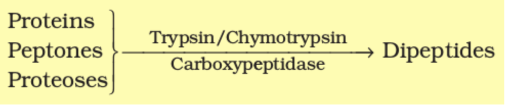
- Proteolytic Enzymes:
- Trypsin and Chymotrypsin: Break proteins into smaller peptides.
- Carboxypeptidase: Further splits peptides into amino acids.
Endocrine Functions:
- The endocrine portion consists of clusters of cells called islets of Langerhans, which secrete hormones:
- Insulin: Lowers blood glucose levels by facilitating glucose uptake.
- Glucagon: Raises blood glucose levels by stimulating glycogen breakdown.
Role of Pancreatic Secretions:
- Pancreatic juice is released into the duodenum via the pancreatic duct.
- Works in concert with bile to digest carbohydrates, proteins, and fats.
Digestion of Food
The process of digestion involves the mechanical and chemical breakdown of complex food molecules into simpler substances that can be absorbed and utilized by the body. This intricate process occurs in sequential steps along the alimentary canal and is facilitated by various enzymes, secretions, and movements of the digestive tract.
Digestion in the Buccal Cavity
The buccal cavity initiates digestion through mechanical and chemical processes:
- Mastication:
- Teeth grind, crush, and chew food into smaller particles, increasing the surface area for enzyme action.
- The tongue aids in manipulating and mixing food with saliva.
- Formation of Bolus:
- Mucus in saliva lubricates and adheres food particles, forming a cohesive mass called the bolus.
- Deglutition (Swallowing):
- The bolus is moved into the pharynx and then into the oesophagus via peristalsis (rhythmic muscular contractions).
Chemical Digestion:
- Saliva, secreted by salivary glands, contains:
- Salivary Amylase (Ptyalin): Hydrolyzes starch (30%) into maltose and dextrins at an optimal pH of 6.8.
- Lysozyme: Acts as an antibacterial agent, protecting the oral cavity from infections.

Digestion in the Stomach
The food is retained for 4–5 hours and mixed thoroughly with acidic gastric juice by the churning action of the stomach’s muscular walls. This partially digested, semi-liquid mass is called chyme.
Gastric Glands: The gastric glands in the stomach mucosa contain three types of specialized cells:
- Mucus Neck Cells:
- Secrete mucus, which protects the stomach lining from the corrosive action of hydrochloric acid (HCl) and lubricates the chyme.
- Chief (Peptic) Cells:
- Produce the proenzyme pepsinogen, which is activated by HCl into pepsin.
- Pepsin, a proteolytic enzyme, breaks proteins into proteoses and peptones (smaller peptide fragments).
- In infants, rennin aids in coagulating milk proteins for digestion.
- Parietal (Oxyntic) Cells:
- Secrete HCl, which maintains an acidic environment (pH 1.8) optimal for pepsin activity.
- Secrete intrinsic factor, essential for the absorption of vitamin B12 in the small intestine.
Additional Secretions:
- Gastric juice also contains small amounts of gastric lipase, which plays a minor role in fat digestion.
Digestion in the Small Intestine
The small intestine is the primary site of chemical digestion and nutrient absorption. Various secretions such as bile, pancreatic juice, and intestinal juice work synergistically to complete the breakdown of biomolecules.
Movements in the Small Intestine:
- Segmental contractions mix the chyme with digestive secretions, while peristalsis propels it forward.
Secretions in the Small Intestine:
- Bile (produced by the liver and stored in the gall bladder):
- Contains bile salts, which emulsify fats into smaller droplets (micelles) to increase enzyme efficiency.
- Bilirubin and biliverdin (bile pigments) are excretory products of hemoglobin breakdown.
- Bile facilitates the activation of lipases.
- Pancreatic Juice (from the pancreas):
- Released into the duodenum via the hepato-pancreatic duct.
- Contains inactive enzymes:
- Trypsinogen, activated by enterokinase (from intestinal mucosa), converts into trypsin, which activates other enzymes such as:
- Chymotrypsinogen → Chymotrypsin.
- Procarboxypeptidases → Carboxypeptidase.
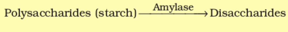
- Amylase: Converts carbohydrates into disaccharides.
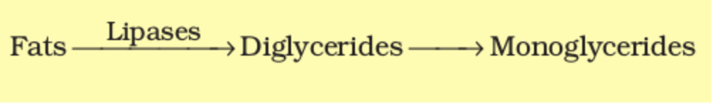
- Lipase: Hydrolyzes triglycerides into glycerol and fatty acids.
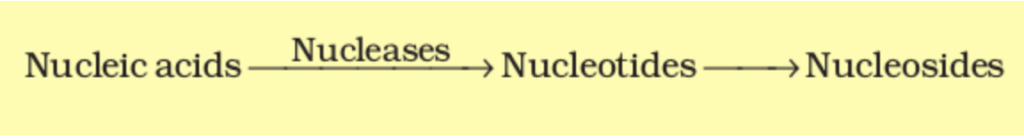
- Nucleases: Digest DNA and RNA into nucleotides.
- Trypsinogen, activated by enterokinase (from intestinal mucosa), converts into trypsin, which activates other enzymes such as:
- Intestinal Juice (Succus Entericus):
- Secreted by goblet cells and brush border cells of the intestinal mucosa.
- Contains enzymes such as:
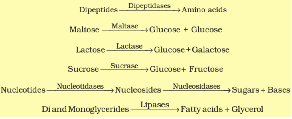
- Disaccharidases (e.g., maltase): Break disaccharides into monosaccharides.
- Dipeptidases: Split dipeptides into amino acids.
- Lipases: Hydrolyze lipids into glycerol and fatty acids.
- Nucleosidases: Break nucleotides into nitrogenous bases, sugars, and phosphates.
pH Regulation:
- Mucus and bicarbonates from the pancreas neutralize the acidic chyme, providing an alkaline pH (7.8) suitable for enzyme activity.
Absorption and Functions of the Large Intestine
The undigested and unabsorbed food material enters the large intestine through the ileocecal valve. While no significant digestive activity occurs here, it performs the following functions:
- Water and Electrolyte Absorption:
- Reabsorbs water, minerals, and certain drugs.
- Mucus Secretion:
- Mucus secreted by goblet cells aids in binding undigested particles and lubricating them for easy passage.
- Formation and Storage of Feces:
- The undigested material, termed feces, is temporarily stored in the rectum and eliminated via the anus during defecation.
Neural and Hormonal Regulation of Digestion
The activities of the gastrointestinal tract are finely regulated by neural and hormonal mechanisms:
- Neural Regulation:
- The sight, smell, or thought of food stimulates the secretion of saliva and gastric juices via reflexes.
- The enteric nervous system and central nervous system (CNS) coordinate muscular contractions and secretory activities.
- Hormonal Regulation:
- Local hormones secreted by the gastric and intestinal mucosa (e.g., gastrin, secretin, cholecystokinin) regulate the production and release of digestive juices.
The enzymes present in succus entericus (intestinal juice) play a crucial role in the final stages of digestion by breaking down complex molecules into their simplest, absorbable forms. These processes occur in close proximity to the mucosal epithelial cells of the small intestine, particularly on the brush border of the intestinal villi.
Enzymatic Actions in Succus Entericus
- Disaccharidases:
- Convert disaccharides into monosaccharides:
- Maltase: Hydrolyzes maltose into two glucose molecules.
- Lactase: Breaks down lactose into glucose and galactose.
- Sucrase: Splits sucrose into glucose and fructose.
- Convert disaccharides into monosaccharides:
- Dipeptidases:
- Hydrolyze dipeptides into individual amino acids.
- Lipases:
- Act on diglycerides and monoglycerides to produce glycerol and fatty acids.
- Nucleosidases and Phosphatases:
- Degrade nucleotides into their components:
- Nitrogenous bases.
- Pentose sugars.
- Phosphates.
- Degrade nucleotides into their components:
Absorption of Digested Products
Absorption refers to the transfer of the end products of digestion across the intestinal mucosa into the bloodstream or lymphatic system. This process is mediated by various transport mechanisms, including passive diffusion, facilitated diffusion, and active transport, depending on the properties of the nutrient.
Mechanisms of Absorption
- Passive Diffusion:
- Substances move along their concentration gradient without requiring energy.
- Examples: Small amounts of monosaccharides (e.g., glucose), amino acids, and chloride ions.
- Facilitated Transport:
- Nutrients are transported with the help of carrier proteins, enabling substances to move down their concentration gradient more efficiently.
- Examples: Glucose and amino acids.
- Active Transport:
- Substances are transported against their concentration gradient, requiring energy (ATP).
- Examples: Monosaccharides, amino acids, and electrolytes like Na⁺.
- Osmosis:
- Water absorption occurs due to osmotic gradients created by solute concentrations in the intestinal lumen and surrounding tissues.
- Fat Absorption:
- Fatty acids and glycerol are insoluble in water and cannot directly enter the blood. Instead:
- They are emulsified into small droplets called micelles by bile salts.
- Micelles diffuse into the intestinal mucosa.
- Inside mucosal cells, they are reassembled into chylomicrons (protein-coated fat globules).
- Chylomicrons are transported into the lacteals (lymphatic vessels in the villi).
- From the lacteals, they enter the bloodstream via the lymphatic system.
- Fatty acids and glycerol are insoluble in water and cannot directly enter the blood. Instead:
Sites of Absorption
- Mouth:
- Limited absorption of substances like certain drugs and alcohol.
- Stomach:
- Absorption of water, alcohol, and some lipid-soluble substances.
- Small Intestine:
- The primary site for nutrient absorption due to its vast surface area (villi and microvilli).
- Absorbs:
- Monosaccharides (e.g., glucose, fructose).
- Amino acids and dipeptides.
- Fatty acids and glycerol.
- Vitamins (e.g., B-complex, C, and fat-soluble vitamins A, D, E, K).
- Minerals (e.g., Na⁺, K⁺, Ca²⁺).
- Water.
- Large Intestine:
- Absorbs:
- Water (reclaiming about 90% of the remaining fluid).
- Some electrolytes (e.g., Na⁺ and Cl⁻).
- Certain vitamins produced by gut microbiota (e.g., Vitamin K, biotin).
- Absorbs:
| Nutrient | Form Absorbed | Transport Mechanism | Site of Absorption |
| Carbohydrates | Monosaccharides (e.g., glucose, fructose) | Active/facilitated transport | Small intestine |
| Proteins | Amino acids, dipeptides | Active/facilitated transport | Small intestine |
| Fats | Fatty acids, glycerol | Diffusion (micelles → chylomicrons → lacteals) | Small intestine |
| Water | Water | Osmosis | Stomach, small intestine, large intestine |
| Electrolytes | Na⁺, Cl⁻, K⁺, etc. | Active/passive transport | Small and large intestines |
| Vitamins | Fat-soluble (A, D, E, K); Water-soluble (B-complex, C) | Diffusion | Small intestine (large intestine for some vitamins) |
| Minerals | Ca²⁺, Mg²⁺, Fe²⁺, etc. | Active transport | Small intestine |
Disorders of the Digestive System
- Inflammation of the Intestinal Tract:
- Cause: Bacterial or viral infections, as well as parasitic infestations (e.g., tapeworm, roundworm, hookworm, pinworm).
- Symptoms: Abdominal pain, cramps, diarrhea, and bloating.
- Treatment: Antibiotics, antiparasitic medications, and rehydration.
- Jaundice:
- Cause: Liver dysfunction leading to the accumulation of bilirubin, a bile pigment, in the blood.
- Symptoms: Yellowing of the skin and eyes.
- Treatment: Treating the underlying liver disease, such as hepatitis or cirrhosis, and managing bile duct blockages.
- Vomiting:
- Cause: Various factors including infections, food poisoning, motion sickness, or psychological triggers.
- Mechanism: Controlled by the vomit center in the medulla oblongata of the brain.
- Symptoms: Ejection of stomach contents through the mouth, often preceded by nausea.
- Treatment: Antiemetic medications to control nausea, hydration, and electrolyte replacement.
- Diarrhea:
- Cause: Infections, food intolerance, inflammatory bowel disease, or certain medications.
- Symptoms: Increased frequency of bowel movements and liquid stools.
- Impact: Decreases absorption of water, nutrients, and electrolytes.
- Treatment: Rehydration therapy (oral rehydration salts), antibiotics for bacterial causes, and dietary adjustments.
- Constipation:
- Cause: Irregular bowel movements, inadequate fiber intake, dehydration, or certain medications.
- Symptoms: Difficulty passing stools, infrequent bowel movements, and feeling of incomplete evacuation.
- Treatment: Increased fiber intake, proper hydration, regular physical activity, and sometimes the use of laxatives.
- Indigestion (Dyspepsia):
- Cause: Inadequate secretion of digestive enzymes, overeating, anxiety, or consumption of spicy or fatty foods.
- Symptoms: Feeling of fullness, bloating, discomfort, or pain in the upper abdomen.
- Treatment: Antacids, dietary changes, reducing stress, and addressing the underlying cause (e.g., enzyme supplements).
FAQ (Previous year questions)
Shree Anna is a term used in India to refer to millets, a group of small-seeded cereal grains known for their high nutritional value and climate resilience.
Examples: Jowar (sorghum), bajra (pearl millet), ragi (finger millet), foxtail millet, proso millet, kodo millet, little millet, and barnyard millet.
These are called Nutri-Cereals because they offer a wide range of health benefits that contribute to overall well-being:
Rich in Nutrients: Millets are packed with essential nutrients such as protein, dietary fiber, iron, calcium, zinc, and B-complex vitamins, making them a wholesome and energy-rich food.
Low Glycemic Index: They release glucose slowly into the bloodstream, preventing sudden spikes in blood sugar levels and making them especially beneficial for people with diabetes.
Gluten-Free: Being naturally gluten-free, millets are an excellent dietary choice for individuals with gluten intolerance or celiac disease.
High Fiber Content: Their high fiber helps improve digestion, prevents constipation, and promotes a healthy gut microbiome.
Heart-Healthy: Regular consumption of millets helps reduce bad cholesterol (LDL) levels and supports better heart health by maintaining blood pressure and vascular function.
Rich in Antioxidants: Millets are a good source of antioxidants, which combat oxidative stress and help strengthen the immune system.
Good for Weight Management: Their high satiety value keeps one full for longer, reducing unnecessary snacking and aiding in weight control.
Suitable for All Ages: Millets are safe and beneficial for all age groups—from growing children to the elderly—due to their comprehensive nutritional profile and easy digestibility.
Efforts to promote Shri Anna –
Renaming as “Shree Anna”: To honor millets’ cultural and nutritional value.
International Year of Millets 2023: India led the global campaign with active promotion.
Support to Millet Farmers: Through schemes like Millet Mission and PKVY.
Promotion in G20 Events: Millets featured as a model for sustainable food systems.
India has been working to position itself as the “Global Hub for Millets”, promoting exports and rese
