
The Cell is the basic structural and functional unit of all living organisms. In Biology, it is considered the building block of life, as all life forms are made up of one or more cells. Cells carry out essential functions that keep an organism alive and functioning.
The Cell
- All organisms are composed of cells.
- Unicellular organisms:
- Made of a single cell.
- Capable of independent existence and performing all essential life functions.
- Multicellular organisms:
- Composed of many cells.
- Cell:
- Fundamental structural and functional unit of life.
- Anything less than a cell cannot lead an independent life.
Cell Theory
- Anton von Leeuwenhoek: First to observe and describe a live cell.
- Robert Brown: Discovered the nucleus.
- Development of the microscope (including the electron microscope) revealed cellular structures in detail.
- Main principles of cell theory:
- All living organisms are composed of cells and their products.
- All cells arise from pre-existing cells.
Cell Structure and Types
General Features:
- Outer membrane: Surrounds the cell and separates it from its surroundings.
- Nucleus:
- Membrane-bound dense structure.
- Contains chromosomes with genetic material (DNA).
- Cytoplasm:
- Semi-fluid matrix filling the cell.
- Site of cellular activities and chemical reactions
Types of Cells:
- Prokaryotic cells and Eukaryotic cells {Include Plant and Animal Cells}
Differences Between Prokaryotic and Eukaryotic Cells
| Feature | Prokaryotic Cell | Eukaryotic Cell |
| Nucleus | No true nucleus (nucleoid region present). | True nucleus surrounded by a nuclear membrane. |
| Size | Small (1-10 µm). | Larger (10-100 µm). |
| Organelles | No membrane-bound organelles (e.g., no mitochondria or ER). | Membrane-bound organelles (e.g., mitochondria, ER, Golgi). |
| DNA | Circular DNA without histones. | Linear DNA associated with histones. |
| Cell Division | Binary fission. | Mitosis and meiosis. |
| Examples | Bacteria, Archaea. | Plants, Animals, Fungi, Protists. |
| Ribosomes | Smaller (70S). | Larger (80S) in the cytoplasm. |
| Cell Wall | Present (made of peptidoglycan in bacteria). | Present in plants (cellulose) and fungi (chitin); absent in animals. |
| Flagella | Simple, made of flagellin. | Complex, made of microtubules. |
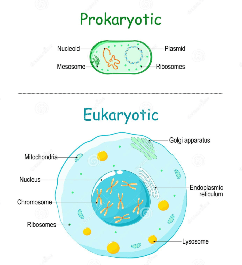
Components of Prokaryotic Cells
- Plasma membrane:
- Outer protective layer of phospholipids.
- Separates the cell from its surroundings.
- Cytoplasm:
- Jelly-like substance inside the cell.
- Suspends cell components.
- DNA:
- Circular genetic material.
- Directs protein creation and cell actions.
- Ribosomes:
- Non-membrane-bound organelles.
- Site of protein synthesis.
- Locomotion structures (in some prokaryotes):
- Cilia and flagella aid movement.
Components of Eukaryotic Cells
- Organised nucleus:
- Enclosed by a nuclear envelope.
- Contains chromosomes with DNA.
- Cytoplasm:
- Highly compartmentalised.
- Contains membrane-bound organelles.
- Membrane-bound organelles:
- Endoplasmic reticulum (ER): Transport of materials.
- Golgi complex: Processing and packaging of substances.
- Lysosomes: Digestion and waste removal.
- Mitochondria: Energy production.
- Microbodies: Various metabolic functions.
- Vacuoles: Storage of substances.
- Non-membrane-bound organelles:
- Ribosomes: Found in the cytoplasm and on rough ER.
- Present in all cells (both prokaryotic and eukaryotic).
- Found in mitochondria and chloroplasts.
- Centrosome (in animal cells): Aids in cell division.
Differences between Plant cell and Animal cell
| Characteristic | Plant Cell | Animal Cell |
| Cell Wall | Present, made of cellulose | Absent |
| Chloroplasts | Present, for photosynthesis | Absent |
| Vacuole | Large, central vacuole | Small or absent |
| Energy Storage | Starch | Glycogen |
| Centrioles | Absent (in most plants) | Present |
| Mobility Structures | Rarely has flagella or cilia | May have flagella (sperm) or cilia |
| Cytokinesis | Formation of a cell plate | Formation of a cleavage furrow |
| Pigments | Chlorophyll and other photosynthetic pigments | Melanin and other functional pigments |
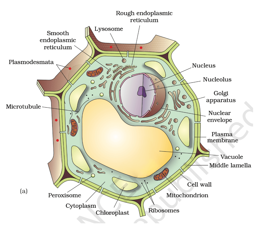
(a) Plant cell
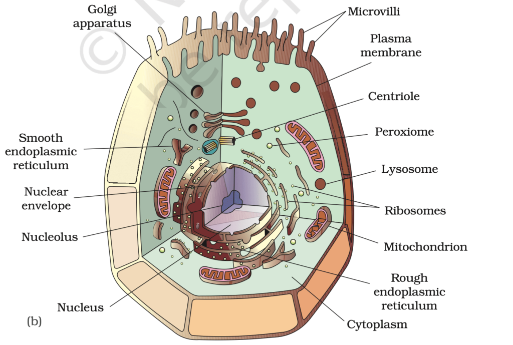
(b) Animal cell
Cell structure and functions
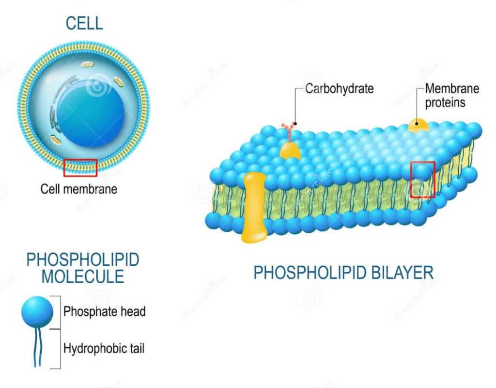
Cell membrane
Structure of Plasma Membrane
- Scientists deduced that the plasma membrane is primarily made up of:
- Lipids (majorly phospholipids).
- Arranged in a bilayer.
- Polar (hydrophilic) heads: Oriented outward
- Nonpolar (hydrophobic) tails: Oriented inward
- Cholesterol: Found between phospholipid molecules.
- Proteins (various functional types).
- Carbohydrates (associated with lipids and proteins).
- Present as glycoproteins and glycolipids.
- Located on the extracellular side of the membrane.
- Lipids (majorly phospholipids).
Functions of Plasma Membrane:-
- Boundary & Protection: Separates the cell from the environment and safeguards internal components.
- Selective Permeability: Controls entry and exit of molecules; maintains homeostasis.
- Transport: Controls the entry and exit of ions, nutrients, and waste.
- Passive: No energy needed (e.g., diffusion of Oxygen and carbon dioxide, osmosis).
- Active: Uses ATP to move molecules against the gradient (e.g., Na+/K+ pump).
- Endocytosis & Exocytosis:
- Endocytosis: Engulfs particles (e.g., phagocytosis for solids).
- Exocytosis: Expels waste or secretions.
- Cell Signaling: Proteins and glycoproteins act as receptors for hormones and other signaling molecules. It helps the cell respond to environmental changes.
- Cell Adhesion: Glycoproteins aid in cell-to-cell attachment. It is essential in tissue formation and stability.
- Energy Conversion: Aids in ATP production in some cells (e.g., prokaryotes).
- Structural Support: Maintains cell shape by interacting with the cytoskeleton.
Cell Wall
Structure and Functions
- The cell wall is a rigid, non-living layer in plants, fungi, algae, and some bacteria. It provides support, protection, and regulates substance movement.
Composition of the Cell Wall:
- In Plants:
- Cellulose: Main structural component.
- Hemicellulose and Pectin: Provide flexibility and structure.
- In Fungi:
- Chitin: Main structural component.
- Glucans: Support the cell wall.
- In Bacteria:
- Peptidoglycan: Main structural component (in Gram-positive and Gram-negative bacteria).
- In Algae:
- Cellulose, Pectin, or Algal-specific polysaccharides: Varies by species.
Endomembrane System
The Endomembrane System is a network of membranous organelles in eukaryotic cells that work together to synthesize, modify, transport, and break down molecules. The key components are:
- Endoplasmic Reticulum (ER): Involved in protein and lipid synthesis.
- Golgi Complex: Modifies, sorts, and packages proteins and lipids.
- Lysosomes: Contain enzymes for digestion and waste breakdown.
- Vacuoles: Store nutrients, waste, and help in cellular homeostasis.
Organelles like mitochondria, chloroplasts, and peroxisomes are not part of this system.
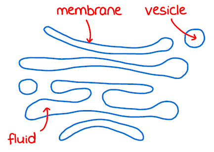
The Endoplasmic Reticulum (ER):
The endoplasmic reticulum (ER) is an important cellular organelle that plays a key role in the synthesis and transport of proteins and lipids. It is a network of tubular structures or membranous sacs found throughout the cytoplasm in eukaryotic cells. The ER divides the intracellular space into two distinct compartments:
- Luminal (inside ER)
- Extra-luminal (cytoplasm)
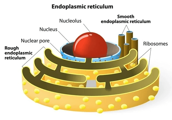
Types of Endoplasmic Reticulum (ER):
- Rough Endoplasmic Reticulum (RER):
- Structure: Studded with ribosomes, giving it a rough appearance.
- Function: Synthesizes proteins for secretion, plasma membrane, or lysosomes.
- Presence: Found in cells that actively produce proteins, like pancreatic cells and plasma cells.
- Smooth Endoplasmic Reticulum (SER):
- Structure: Lacks ribosomes, giving it a smooth appearance.
- Function: Synthesizes lipids (e.g., steroidal hormones), detoxifies drugs, and metabolizes carbohydrates. It stores and regulates calcium in Muscle Cells.
Golgi apparatus (Golgi Bodies):
- Discovery: First observed by Camillo Golgi in 1898, these densely stained structures near the nucleus were named after him.
- Structure: Golgi bodies consist of flattened, disc-shaped sacs called cisternae (0.5 µm to 1.0 µm in diameter). These are stacked parallel to each other.
- Function: The Golgi apparatus is responsible for modifying, sorting, and packaging proteins and lipids from the endoplasmic reticulum (ER) for delivery to other parts of the cell or outside the cell. ⇒ “cell’s post office”
Lysosomes
- Structure: Membrane-bound vesicles formed by the Golgi apparatus.
- Content: Contain hydrolytic enzymes (hydrolases), including:
- Lipases: Break down lipids.
- Proteases: Break down proteins.
- Carbohydrases: Break down carbohydrates. These enzymes function best in an acidic environment.
- Function: Responsible for digesting carbohydrates, proteins, lipids, and nucleic acids.
- Role: Key in cellular digestion, waste removal, and autophagy (recycling damaged cellular components). ⇒ Cell’s “garbage disposal” system.
Vacuoles
- A vacuole is a membrane-bound sacs found in the cytoplasm that stores water, nutrients, waste, and other substances.
- In plant cells, the large central vacuole helps maintain cell shape by holding water and creating turgor pressure.
- Vacuoles also play a role in waste disposal and maintaining the cell’s internal balance.
- Animal cells have smaller vacuoles, mostly for storage and transport.
Mitochondria:
- Structure:
- Mitochondria are double-membrane-bound organelles, typically sausage-shaped or cylindrical in form, with a diameter of 0.2–1.0 µm and a length of 1.0–4.1 µm.
- The outer membrane forms the boundary of the organelle, while the inner membrane is folded into cristae(increased surface area).
- The inner compartment contains a dense substance known as the matrix, which houses mitochondrial DNA, ribosomes (70S), and enzymes required for protein synthesis.
- Function:
- Mitochondria are the powerhouses of the cell, involved in aerobic respiration to produce ATP (adenosine triphosphate), the energy currency of the cell.
- They contain their own circular DNA and can divide by fission.
Plastids
- Structure: Plastids are large, membrane-bound organelles found in plant cells and some euglenoids. They contain various pigments that impart specific colors to plants.
- Types of Plastids:
- Chloroplasts: Contain chlorophyll and carotenoid pigments.
- Responsible for photosynthesis by capturing light energy.
- Chromoplasts: Contain carotenoid pigments such as carotene and xanthophyll.
- Impart yellow, orange, or red colors to plant parts.
- Leucoplasts: Colorless plastids that store starch, oils, and proteins.
- Chloroplasts: Contain chlorophyll and carotenoid pigments.
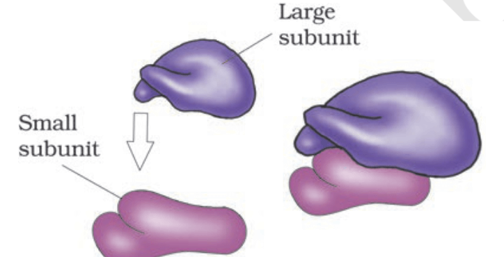
Ribosomes
Ribosomes are crucial molecular machines in cells responsible for protein synthesis (translation). They play a central role in translating genetic information encoded in messenger RNA (mRNA) into functional proteins.
- Structure of Ribosomes
- Composition: Made up of ribosomal RNA (rRNA) and proteins.
- Subunits:
- Larger Subunit: Forms peptide bonds between amino acids.
- Smaller Subunit: Reads mRNA and guides protein synthesis.
- Svedberg Unit (S): Ribosomal subunits are measured in Svedberg units, indicating size and density.
- Location:
- Free Ribosomes: Floating in the cytoplasm, synthesizing proteins for the cytosol.
- Bound Ribosomes: Attached to the rough ER, synthesizing proteins for membranes, organelles, or secretion.
Function of Ribosomes
- Protein Synthesis: Ribosomes are the sites of protein synthesis in the cell. This process, called translation, involves decoding the genetic instructions carried by messenger RNA (mRNA) to assemble amino acids into polypeptide chains (proteins).
Ribosome and Genetic Code:
- Ribosomes read mRNA codons (coding for a specific amino acid )and tRNA brings the corresponding amino acids.
Prokaryotic vs. Eukaryotic Ribosomes:
- Prokaryotes: Smaller (70S), free in cytoplasm.
- Eukaryotes: Larger (80S), free or attached to the ER.
- Despite size differences, ribosomes in both types of cells serve the same function. The structural differences allow antibiotics to target bacterial ribosomes without affecting eukaryotic cells.
Cytoskeleton
The cytoskeleton is a network of protein fibers in the cell that provides structure, support, and shape. It is composed of:
- Microfilaments: Thin fibers that support its shape and enable its movement (e.g., during muscle contraction).
- Intermediate Filaments: Provide mechanical support and help maintain the cell’s integrity.
- Microtubules: Hollow tubes that help with cell shape, transport, and cell division (they form the spindle fibers during mitosis).
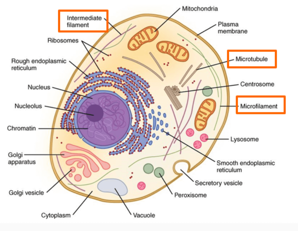
Centrosome and Centrioles
- Centrosome: The centrosome is the region in the cell that organizes microtubules and plays a key role in cell division. It consists of a pair of centrioles and surrounding pericentriolar material.
- Centrioles: Centrioles are cylindrical structures made of microtubules. In animal cells, they help organize the spindle fibers during mitosis and meiosis, ensuring proper chromosome separation during cell division.
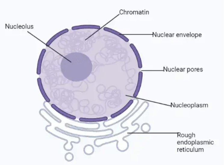
The Nucleus: Structure and Functions
Structure of the Nucleus:
- The nucleus is a membrane-bound organelle found in eukaryotic cells.
- It is surrounded by a double-layered membrane called the nuclear envelope, which has pores for material exchange between the nucleus and cytoplasm.
- Inside the nucleus is a substance called nucleoplasm (Nuclear Matrix), which contains chromatin (DNA and proteins) and the nucleolus (site of ribosome production).
Functions of the Nucleus:
- Genetic Material Storage: The nucleus stores DNA, which contains the genetic instructions for the cell.
- Gene Expression: It regulates gene expression by controlling which genes are turned on or off.
- Cell Division: The nucleus plays a key role in cell division, ensuring accurate replication and distribution of genetic material.
- Ribosome Production: The nucleolus within the nucleus synthesizes ribosomal RNA (rRNA) and assembles ribosomal subunits.
Discovery and Naming
- 1831: Robert Brown first described the nucleus.
- Chromatin: The material in the nucleus that stains with basic dyes was named by Flemming.
Variations in the Nucleus
Most cells have a single nucleus, but there are exceptions:
- Multinucleated Cells: Found in organisms like fungi, some algae, and skeletal muscle cells.
- Enucleated Cells: Mature mammalian red blood cells (e.g., erythrocytes) and sieve tube cells in vascular plants lack a nucleus.
Chromatin is a complex of DNA and proteins (mainly histones) found in the nucleus.
- It packages DNA into a compact structure to fit inside the nucleus.
- Controls gene expression by regulating access to DNA.
- During cell division, chromatin condenses to form chromosomes.
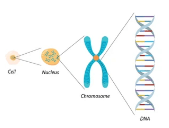
Chromosomes
During cell division, chromatin condenses to form structured chromosomes that are visible under a microscope.
Chromosome Composition
- Made of:
- DNA: The genetic material.
- Proteins: Histones and non-histones that help in DNA organization.
- In humans:
- Each cell contains 46 chromosomes.
- The total length of DNA in a single human cell is approximately 2 meters.
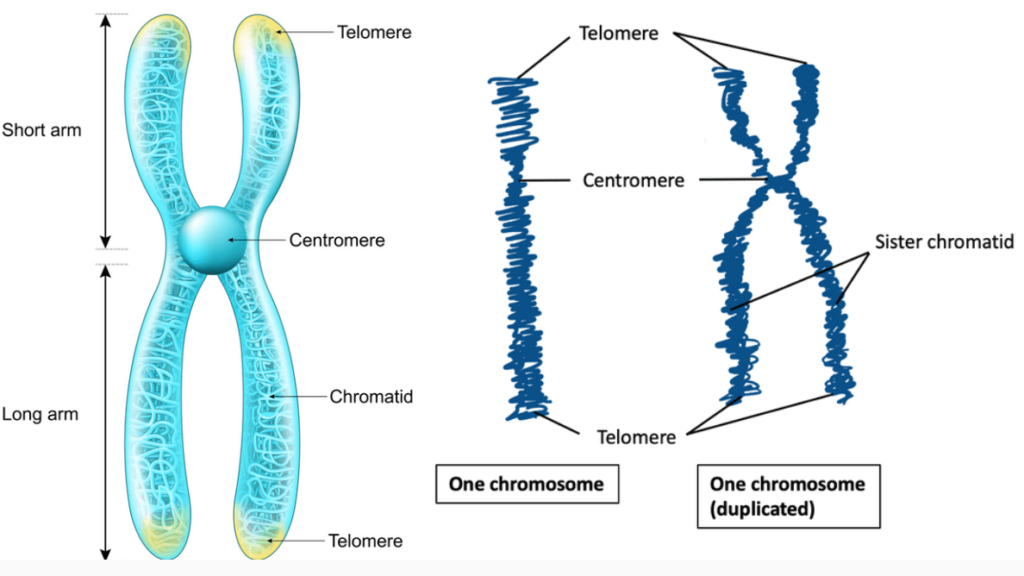
Chromosome Structure
- Centromere: A primary constriction that holds two sister chromatids together.
- Kinetochore: Disc-shaped structures on either side of the centromere, where spindle fibers attach during cell division.
Microbodies
- Membrane-bound vesicles found in plant and animal cells.
- Contain enzymes that perform specific functions like detoxification and lipid metabolism.
CELL CYCLE
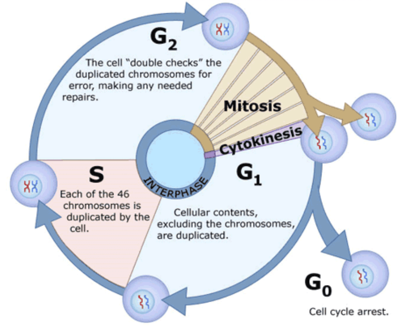
Cell division is a fundamental process in all living organisms, allowing growth, repair, and reproduction. It ensures that genetic material is accurately replicated and distributed to progeny cells. The process involves a series of well-coordinated events: DNA replication, cell growth, and division. These events are collectively organized into a sequence called the cell cycle.
Thus, The cell cycle refers to the series of events a cell goes through as it grows, duplicates its DNA, and divides to form two daughter cells.
Phases of the Cell Cycle
The cell cycle is divided into two main phases:
- Interphase (Preparation phase): The preparation phase, during which the cell grows and replicates its DNA. It accounts for about 95% of the total cell cycle duration in human cells.
- G1 Phase: Cell grows and performs normal functions.
- S Phase: DNA is replicated.
- G2 Phase: Cell prepares for division by growing and checking for errors.
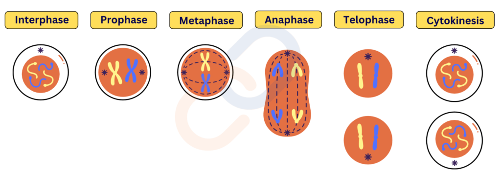
- M Phase (Mitotic Phase →Division phase): The actual division phase, involving the division of the nucleus (karyokinesis) and cytoplasm (cytokinesis).
Mitosis : Division of the nucleus into two identical nuclei.
- Prophase:
- Chromatin condenses into chromosomes.
- The nuclear membrane begins to break down.
- Spindle fibers start forming.
- Metaphase:
- Chromosomes align in the center of the cell (at the metaphase plate).
- Spindle fibers attach to the centromeres of chromosomes.
- Anaphase:
- Sister chromatids are pulled apart to opposite poles of the cell by spindle fibers.
- Telophase:
- Chromatids reach opposite poles and decondense into chromatin.
- The nuclear membrane reforms around each set of chromosomes.
Cytokinesis:
- The cytoplasm divides, resulting in two separate daughter cells, each with its own nucleus.
- Animal cells: A cleavage furrow forms, pinching the cell into two.
- Plant cells: A cell plate forms at the center, developing into a new cell wall.
Duration of Cell Cycle
- The time required for a cell to complete one cycle varies by organism and its type.
- Human cells: ~24 hours.
- Interphase: ~23 hours.
- M Phase: ~1 hour.
- Yeast cells: ~90 minutes.
Mitosis Vs Meiosis
Mitosis
- Mitosis is the type of cell division responsible for growth, tissue repair, and asexual reproduction.
- It results in two genetically identical daughter cells with the same chromosome number as the parent cell.
Meiosis :
- Meiosis is a specialized cell division that reduces the chromosome number by half, producing four haploid cells (gametes like sperm and eggs) from a single diploid cell.
- It occurs in two stages, Meiosis I and Meiosis II, and is essential for sexual reproduction.
- Importance:
- Meiosis ensures genetic variation in offspring.
- It is crucial for maintaining a stable chromosome number across generations.
Key Features of Meiosis:
- Reduces chromosome number from diploid (2n) to haploid (n).
- Ensures genetic diversity through crossing over and independent assortment.
- Produces gametes (sperm and egg cells) in animals and spores in plants.
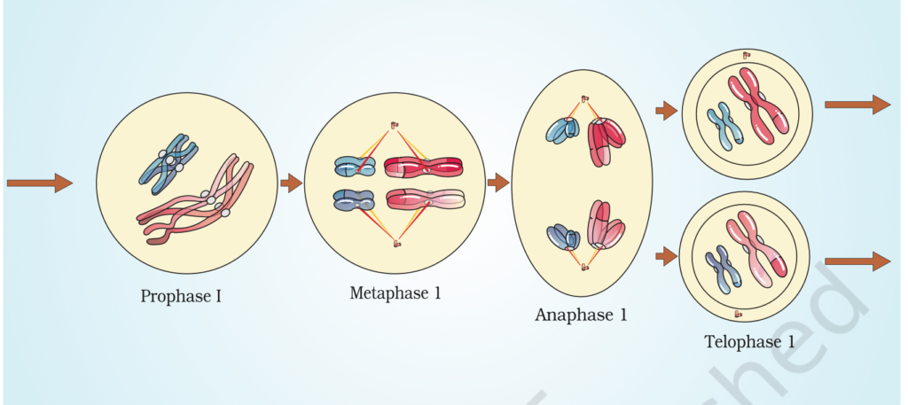
Stages of Meiosis:
- Meiosis I (Reductional Division): The first division reduces the chromosome number from diploid (2n) to haploid (n).
- Prophase I: Homologous chromosomes pair up, and crossing over occurs (exchange of genetic material).
- Metaphase I: Homologous chromosome pairs align at the equator.
- Anaphase I: Homologous chromosomes are pulled to opposite poles (chromatids remain attached).
- Telophase I & Cytokinesis: Two haploid cells form, each with half the original chromosome number.
- Interkinesis: A brief resting phase occurs, with no DNA replication
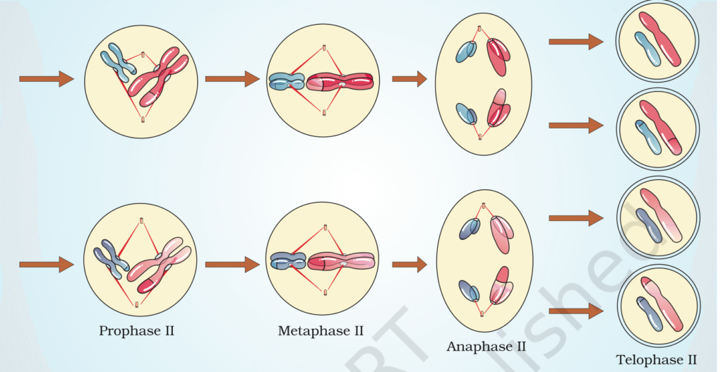
Meiosis II (Equational Division): This division resembles mitosis and separates sister chromatids.
- Prophase II: Chromosomes condense again in both haploid cells.
- Metaphase II: Chromosomes align at the equator.
- Anaphase II: Sister chromatids are pulled apart to opposite poles.
- Telophase II & Cytokinesis: Four haploid daughter cells are formed, each genetically unique.
Differences Between Mitosis and Meiosis
| Aspect | Mitosis | Meiosis |
| Purpose | Growth, repair, and asexual reproduction | Formation of gametes for sexual reproduction |
| Number of Divisions | 1 division | 2 divisions (Meiosis I and Meiosis II) |
| Number of Daughter Cells | 2 daughter cells | 4 daughter cells |
| Chromosome Number | Maintains the same (diploid, 2n → 2n) | Reduces by half (diploid, 2n → haploid, n) |
| Genetic Variation | Daughter cells are identical to the parent cell | Daughter cells are genetically unique |
| Crossing Over | Does not occur | Occurs during Prophase I |
| Type of Cells Produced | Somatic (body) cells | Gametes (sperm or egg cells) |
| Occurrence | In all body cells | Only in reproductive organs (testes/ovaries) |
| Role in Evolution | Does not contribute to genetic variation. | Increases genetic diversity through crossing over and independent assortment. |
| Cytokinesis | Occurs once at the end of telophase, resulting in two cells. | Occurs twice: once after meiosis I and once after meiosis II, resulting in four cells. |
| Significance | * Growth: Allows organisms to grow by producing more cells. * Repair: Replaces damaged or dead cells (e.g., in wounds). * Asexual Reproduction: In single-celled organisms, mitosis is the basis for reproduction. * Maintains Genetic Stability: Produces genetically identical cells, preserving the chromosome number. | * Gamete Formation: Produces sperm and egg cells for sexual reproduction. * Maintains Chromosome Number: Ensures that offspring have the correct chromosome number (haploid gametes combine to form a diploid zygote). * Genetic Variation: Introduces diversity through crossing over and independent assortment, which is important for evolution and adaptation. |
