
Hormones are chemical messengers in the body that regulate various physiological processes such as growth, metabolism, and reproduction. In Biology, they are studied as essential substances secreted by glands that travel through the bloodstream to target organs, ensuring proper functioning and balance in the body.
Hormones
- Hormones are chemical messengers secreted by the glands of the endocrine system. They regulate critical physiological functions by transmitting signals to various organs and tissues, influencing growth, metabolism, reproduction, mood, and other vital body functions.
- The regulation of these hormones is essential for maintaining the body’s internal balance, known as homeostasis.
What Are Hormones?
- Hormones are specialized proteins, peptides, amino acids, or steroid molecules that are produced and released by endocrine glands.
- These hormones enter the bloodstream and travel to target organs and tissues where they bind to specific receptors to trigger various biological responses.
- Hormones are involved in a wide range of bodily functions, including regulating metabolism, controlling growth, balancing the immune system, and managing stress and reproduction.
Functions of Hormones
- Homeostasis: Hormones like ADH and aldosterone maintain the internal environment by regulating water and electrolyte balance.
- Metabolic Regulation: Thyroid hormones and insulin play critical roles in carbohydrate, protein, and lipid metabolism.
- Growth and Development: Growth hormone and sex hormones regulate cellular proliferation, tissue differentiation, and reproductive maturity.
- Stress Response: Adrenal hormones mediate the physiological response to acute and chronic stress.
- Reproductive Function: Gonadal hormones regulate gametogenesis and secondary sexual traits.
- Immune System Modulation: Thymus hormones enhance immune defense, especially in early life.
Types of Hormones:
Peptide Hormones:
- Structure: Composed of chains of amino acids.
- Examples: Insulin, Growth Hormone (GH), Parathyroid Hormone (PTH), Antidiuretic Hormone (ADH).
- Mechanism of Action: They cannot pass through the cell membrane due to their size. Therefore, they bind to receptors on the cell surface, triggering intracellular signaling pathways through second messengers like cAMP.
- Functions: Regulate processes like metabolism (insulin), growth (GH), and water balance (ADH).
Steroid Hormones:
- Structure: Derived from cholesterol.
- Examples: Cortisol, Estrogen, Testosterone, Progesterone, Aldosterone.
- Mechanism of Action: Steroid hormones are lipophilic (fat-soluble) and can easily pass through cell membranes. They bind to intracellular receptors and directly influence gene expression in the nucleus, leading to changes in protein synthesis.
- Functions: Regulate immune response (cortisol), sexual characteristics (estrogen and testosterone), and electrolyte balance (aldosterone).
Amino Acid Derivatives:
- Structure: Derived from the amino acids tyrosine or tryptophan.
- Examples: Thyroxine (T4), Epinephrine (Adrenaline), Norepinephrine (Noradrenaline), Melatonin.
- Mechanism of Action: These hormones may act on cell surface receptors or enter the cell and interact with intracellular receptors.
- Functions: Control metabolism (thyroxine), the body’s fight-or-flight response
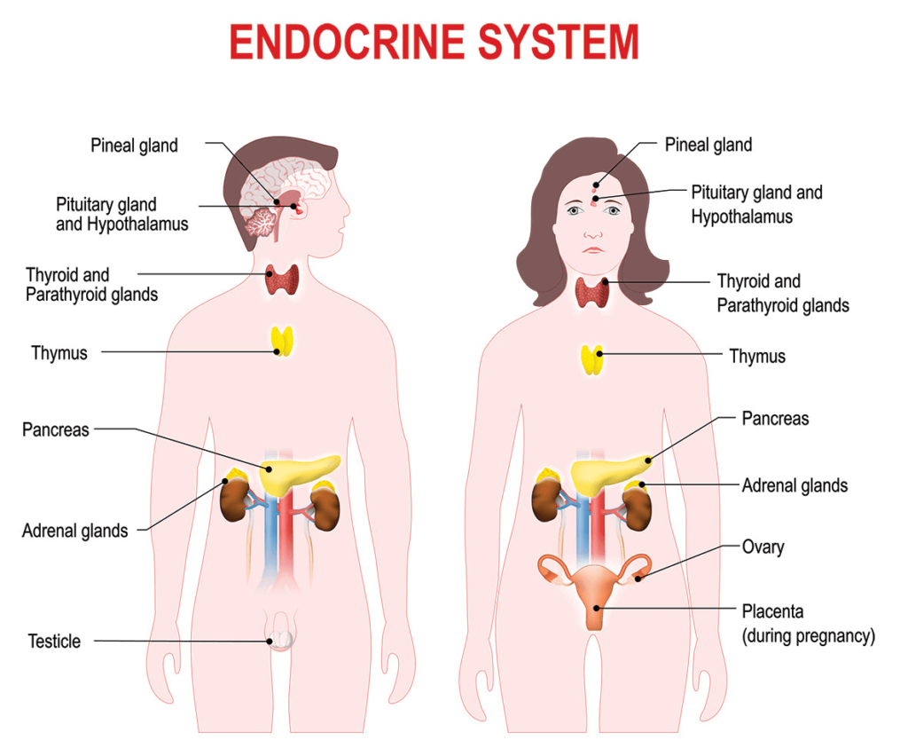
Endocrine glands and hormones
- Endocrine glands are specialized organs in the body that secrete hormones directly into the bloodstream, which then travel to target organs or tissues to regulate various physiological processes.
- Unlike exocrine glands, endocrine glands do not have ducts; they are referred to as ductless glands.
Characteristics of Endocrine Glands
- Ductless Nature: Endocrine glands release hormones directly into the blood rather than through ducts.
- Hormonal Secretion: They produce hormones that regulate specific functions of target cells or organs.
- Vascularization: These glands are richly supplied with blood vessels to facilitate the rapid transport of hormones.
- Target Specificity: Hormones act on specific receptors located in their target cells or organs
1. Hypothalamus
- Located in the brain below the thalamus, forming the floor of the third ventricle.
- Known as the “master of the master gland” because it controls the pituitary gland, which in turn controls other endocrine glands.
- Releases Releasing Hormones or Inhibiting Hormones (Signals to the pituitary gland)
- Releasing Hormones → Stimulate Pituitary to release hormones like:
- Thyroid Stimulating Hormone (TSH) → Stimulates Thyroid (Metabolism)
- Growth Hormone (GH) → Stimulates Growth
- Adrenocorticotropic Hormone (ACTH) → Stimulates Adrenal Glands (Cortisol, Stress)
- Gonadotropins (FSH, LH) → Stimulate Reproductive Organs (Sex hormones)
- Inhibiting Hormones → Stop the release of hormones like:
- Somatostatin → Stops Growth Hormone
- Dopamine → Stops Prolactin (Milk production)
- Oxytocin and Antidiuretic Hormone (ADH): Produced by the hypothalamus but stored and secreted by the posterior pituitary.
- Role in Negative Feedback Loop → When hormone levels are high → Hypothalamus reduces hormone release to keep balance!
- Deficiencies:
- Secondary hypopituitarism, leading to reduced secretion of hormones like GH, ACTH, or TSH.
- Excess:
- Can cause secondary hyperpituitarism, leading to disorders such as gigantism (GH excess) or hyperthyroidism.
2. Pituitary Gland: the Master Gland
- Located: A pea-shaped gland situated at the base of the brain, below the hypothalamus.
- Divided into 3 parts:
- Anterior Pituitary (Front)
- Posterior Pituitary (Back)
- Intermediate Lobe (Produces MSH)
Anterior Pituitary:
- Releases hormones that control other glands and body functions:
- Thyroid Stimulating Hormone (TSH) → Stimulates Thyroid (Regulates metabolism)
- Growth Hormone (GH) → Stimulates Growth of tissues and bones
- Adrenocorticotropic Hormone (ACTH) → Stimulates Adrenal Glands (Cortisol for stress)
- Follicle-Stimulating Hormone (FSH) → Stimulates Ovaries/Testes (Egg/sperm production)
- Luteinizing Hormone (LH) → Stimulates Ovulation/Testes (Sex hormones)
- Prolactin → Stimulates Milk Production in females
- Melanocyte-Stimulating Hormone (MSH) → Stimulates Melanin Production (Skin pigmentation)
Posterior Pituitary:
- Stores and releases hormones made by the Hypothalamus:
- Oxytocin → Uterine contractions (Birth), Milk ejection
- Vasopressin (ADH) → Regulates Water Balance (Kidneys)
- Deficiencies:
- Dwarfism (GH), hypothyroidism (TSH), infertility (LH/FSH), or diabetes insipidus (ADH).
- Excess:
- Acromegaly (GH excess), Cushing’s disease (ACTH excess), or SIADH (ADH excess).
3. Thyroid Gland
- Structure and Location:
- A butterfly-shaped gland located in the neck, anterior to the trachea and inferior to the larynx.
- Consists of two lobes connected by an isthmus.
- Hormones Secreted
- Thyroxine (T4) and Triiodothyronine (T3): Regulate metabolic rate, protein synthesis, and energy production.
- Calcitonin: Lowers blood calcium levels by inhibiting osteoclast activity and promoting calcium storage in bones.
- Iodine is essential for the production of T3 and T4. The thyroid uses iodine from food (like salt and seafood) to produce these hormones.
- Lack of iodine → The thyroid cannot produce enough T3 and T4, leading to problems like goiter (enlarged thyroid).
- Deficiencies:
- Hypothyroidism: Fatigue, weight gain, cold intolerance, goiter.
- In children, it can lead to cretinism, characterized by stunted growth and mental retardation.
- Excess:
- Hyperthyroidism: Weight loss, heat intolerance, irritability (e.g., Grave’s disease).
4. Parathyroid Glands
- Structure and Location:
- Four small, oval glands located on the posterior surface of the thyroid gland.
- Hormones Secreted: Parathyroid Hormone (PTH) → Increases blood calcium levels by:
- Stimulating bone breakdown (releasing calcium from bones into the bloodstream).
- Increasing calcium absorption in the intestines.
- Reducing calcium loss through the kidneys.
- The parathyroid hormone (PTH) works in opposition to calcitonin (released by the thyroid) which lowers blood calcium levels. Together, they maintain calcium homeostasis in the body.
- Deficiencies:
- Hypoparathyroidism: Hypocalcemia, tetany, seizures.
- Excess:
- Hyperparathyroidism: Hypercalcemia, osteoporosis, kidney stones.
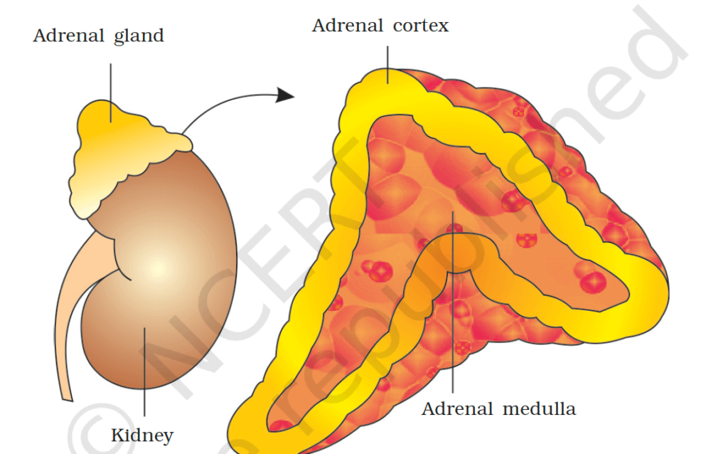
5. Adrenal Glands
- The adrenal glands are two small glands located on top of each kidney.
- Main Function: The adrenal glands produce hormones that help regulate stress response, metabolism, immune function, and salt balance.
Parts of the Adrenal Glands:
- Adrenal Cortex (Outer part): Produces steroid hormones.
- Glucocorticoids (e.g., Cortisol): Help the body respond to stress by regulating glucose metabolism and suppressing inflammation.
- Mineralocorticoids (e.g., Aldosterone): Regulate sodium and potassium balance, thereby influencing blood pressure.
- Androgens: Contribute to the development of secondary sexual characteristics and libido.
- Adrenal Medulla (Inner part)
- Produces Epinephrine (Adrenaline) and Norepinephrine (Noradrenaline)).
- Adrenaline → Increases heart rate, blood pressure, and energy (Stimulates the breakdown of glycogen into glucose) in response to stress (fight or flight).
- Noradrenaline (Norepinephrine) → Known as the emergency hormone, it helps the body react in high-stress situations.
- How the Adrenal Glands Work:
- When the body faces stress, the hypothalamus signals the pituitary gland, which then signals the adrenal glands to release adrenaline and cortisol.
- Cortisol helps manage stress and regulate energy, while adrenaline triggers the “fight or flight” response, preparing the body for quick action.
- Deficiencies:
- Addison’s disease (Deficiency of cortisol and aldosterone): Fatigue, low blood pressure, electrolyte imbalance.
- Excess:
- Cushing’s syndrome (Excess cortisol): Central obesity, hypertension, diabetes.
- Hyperaldosteronism (Excess aldosterone)
6. Pancreas
- Located in the abdomen, posterior to the stomach.
- Mixed gland : Pancreas has both endocrine (2%) and exocrine (98 %) functions:
- Endocrine: Regulates blood sugar levels.
- Exocrine: Produces digestive enzymes to help digest food.
Islets of Langerhans:
- The endocrine part of the pancreas is made up of clusters of cells called Islets of Langerhans, which produce hormones involved in regulating blood sugar.
Types of Cells in the Islets of Langerhans:
- Alpha Cells → Produce Glucagon:
- Function: Raises blood sugar by stimulating the liver to release stored glucose (glycogen).
- Beta Cells → Produce Insulin:
- Function: Lowers blood sugar by helping cells absorb glucose from the bloodstream.
- Delta Cells → Produce Somatostatin:
- Function: Inhibits the release of insulin and glucagon, helping to regulate the balance between blood sugar levels and digestive activity.
- Gamma Cells → Produce Pancreatic Polypeptide:
- Function: Regulates the secretion of digestive enzymes and affects food intake.
How the Pancreas Regulates Blood Sugar:
- When blood sugar is high (after eating), the pancreas releases insulin from beta cells to help cells take in glucose, lowering blood sugar.
- When blood sugar is low (between meals), the pancreas releases glucagon from alpha cells, signaling the liver to release stored glucose, raising blood sugar.
Exocrine Function (Digestive Enzymes):
- The pancreas also produces digestive enzymes (like amylase, lipase, and proteases) that are released into the small intestine to help break down carbohydrates, fats, and proteins.
Excess:
- Hyperinsulinism (Excess insulin): Can lead to hypoglycemia (low blood sugar), causing symptoms like shaking, sweating, and dizziness.
- Insulinoma: Rare benign tumor in the pancreas → Excessive insulin production. It leads to frequent episodes of hypoglycemia.
Deficiency:
- Diabetes Mellitus (Deficiency of insulin or poor insulin function):
- Type 1 Diabetes: The body does not produce enough insulin (due to damaged beta cells).
- Type 2 Diabetes: The body becomes resistant to insulin.
- Symptoms: High blood sugar, fatigue, thirst, frequent urination, and weight loss.
- Hyperglycemia: High blood sugar levels due to insufficient insulin.
- Diabetic Ketoacidosis (DKA): A life-threatening condition in insulin deficiency where the body uses fat for energy, leading to acidic ketone buildup in the blood.
Insulin and its function:
- Produced by: Beta cells of the islets of Langerhans in the pancreas.
- Trigger: Increased blood glucose (after eating).
- Molecular Structure: A protein made up of two peptide chains (A-chain and B-chain) linked by disulfide bonds. Initially, it’s produced as proinsulin, which is then converted into active insulin.
Functions of Insulin
- Regulating Blood Glucose Levels:
- Lowers blood glucose by promoting the uptake of glucose into cells (e.g., muscle and fat cells).
- Stimulates the liver to store glucose as glycogen (glycogenesis).
- Inhibits the formation of new glucose in the liver (gluconeogenesis).
- Energy Storage:
- Promotes glycogen synthesis in the liver and muscles.
- Encourages fat storage in adipose tissues after meals (lipogenesis).
- Cellular Uptake of Glucose:
- Binds to specific receptors on cell surfaces, activating glucose transport proteins (GLUT), allowing glucose to enter cells for energy production or storage.
- Protein Synthesis:
- Stimulates amino acid uptake in muscle tissues.
- Inhibits protein breakdown, promoting growth and repair.
- Regulation of Fat Metabolism:
- Suppresses the breakdown of fats (lipolysis).
- Enhances the formation of fats (lipogenesis).
Regulation of Insulin Secretion
- Feedback Mechanism: Insulin secretion increases when blood glucose levels rise (e.g., after meals) and decreases during fasting.
- Other Stimuli:
- Influenced by neural signals (e.g., during stress/exercise).
- Affected by increased fatty acid levels and hormones like glucagon and growth hormone.
Medical Use of Insulin: Insulin Therapy:
- Used for Type 1 diabetes or advanced Type 2 diabetes.
- Types of insulin: Short-acting, long-acting, or rapid-acting.
7. Gonads
- The gonads are the reproductive glands. In males, they are the testes, and in females, they are the ovaries.
- Main Function:
- The gonads are responsible for producing gametes (sperm in males and eggs in females) and sex hormones(which control secondary sexual characteristics and reproduction).
- Hormones Produced by Gonads:
- In Males (Testes): Testosterone → The primary male sex hormone.
- Function: Responsible for the development of male secondary sexual characteristics such as facial hair, deep voice, muscle growth, and promotes spermatogenesis .
- In Females (Ovaries):
- Estrogen → The primary female sex hormone.
- Function: Regulates the menstrual cycle, promotes the development of female secondary sexual characteristics like breast development, widening of hips, and egg maturation.
- Progesterone → Works with estrogen to regulate the menstrual cycle and pregnancy.
- Function: Prepares the uterus for implantation of a fertilized egg and helps maintain pregnancy.
- Estrogen → The primary female sex hormone.
- Inhibin → Inhibits the production of follicle-stimulating hormone (FSH).
- Function: Regulates the growth and development of ovarian follicles.
- Excess:
- Hyperandrogenism (Excess testosterone in females):
- Symptoms: Excessive hair growth, deepening of the voice, acne.
- Estrogen Dominance (Excess estrogen):
- Symptoms: Heavy periods, mood swings, increased risk of blood clots.
- Polycystic ovarian syndrome (PCOS).
- Hyperandrogenism (Excess testosterone in females):
- Deficiency:
- Hypogonadism (Deficiency of testosterone or estrogen):
- In males: Leads to low libido, decreased muscle mass, and infertility.
- In females: Leads to irregular periods, infertility, and hot flashes.
- Hypogonadism (Deficiency of testosterone or estrogen):
8. Pineal Gland
- The pineal gland is a small, pea-shaped gland located deep in the brain, near the center.
- Hormones Secreted: Melatonin → Regulates sleep patterns and the body’s circadian rhythm (internal body clock).
- Function: Melatonin levels rise in the evening to promote sleep and fall in the morning to help wakefulness. It is influenced by light exposure, particularly daylight and darkness.
- Deficiencies: Insomnia, disrupted sleep patterns.
- Excess: Seasonal Affective Disorder (SAD).
9. Thymus
- The thymus is a small gland located behind the sternum and in front of the heart, mainly in the upper chest.
- Hormones Secreted: Thymosin → Promotes the development and differentiation of T-lymphocytes, critical for adaptive immunity.
- Thymosin helps T cells mature in the thymus and become active in fighting infections and diseases.
- Deficiencies: Thymic Hypoplasia (Underdeveloped thymus) → Weakened immune system.
